From Print to Wool: Vesalius and the ‘Knit your own womb’ Movement
 https://orcid.org/0000-0001-5874-8183
The Open University
helen.king@open.ac.uk
SUMMARY
The womb has been represented in many ways across Western European history, from miracle to sewer. This chapter begins with some of the earliest ways of showing the womb in printed materials. It then looks at what happens when different physical media are used to portray this and other body parts, focusing in particular on the impact of wool and similar materials in making the womb not only more approachable for women but also a potential political tool in claiming women’s rights.
KEYWORDS – body fluids, craft, knitting, uterus, political action
Du dessin au tricot : Vesalius et le mouvement « Tricotez-vous un vagin »
RÉSUMÉ
L’utérus a été représenté de nombreuses façons au cours de l’histoire de l’Europe occidentale, allant du miracle à l’égout. Ce chapitre commence par explorer certaines des premières représentations de l’utérus dans les documents imprimés. Il s’intéresse ensuite à ce qui se passe lorsque différents supports matériels sont utilisés pour représenter cette partie du corps (et d’autres), en mettant particulièrement l’accent sur l’impact de la laine et de matériaux similaires. Ces derniers rendent l’utérus non seulement plus accessible aux femmes, mais en font aussi un outil politique potentiel dans la revendication de leurs droits.
MOTS-CLÉS – fluides corporels, artisanats, tricotage, utérus, action politique
Over the last three decades or so, the history of gynaecology in pre-modern Western Europe has largely focused on the fluids of the body, rather than on its organs. In order to destabilise our assumptions that people at all times and in all places experience their bodies in the same way, it has been important to shift the focus away from what we so easily take for granted. Gail Kern Paster’s 1993 book, The Body Embarrassed: Drama and the Disciplines of Shame in Early Modern England, therefore started from the insight that our apparently fixed, biological bodies can only be understood “in terms of culturally available discourses,” and that for the pre-modern and early modern periods this meant beginning with humoral theory.[1] Today, the four humours, or other fluid models of the body, are too easily regarded as mere intellectual curiosities of the past, but Paster argued that we need to understand that this approach to the body went far beyond metaphor, and that we should try to appreciate “the formative effects of physiological theory on the subject”; that is, the ways in which our deeply-rooted theories about the body affect how we experience being embodied.[2] Our bodies are very different places if, for example, we move away from our own knowledge that menstruation is the result of a hormonally-caused shedding of the womb lining which, in any month, may or may not occur, and instead understand it as an essential by-product of digestion, with the blood accumulating all the time, meaning that any failure of this blood to come out of the body in a regular way is seen as a serious problem.[3] Within humoral medicine, both sexes were understood as composed of fluids, but women were regarded as wetter – in Hippocratic medicine, it was precisely women’s wet and spongy flesh which meant blood was stored and then released – and often also as more “leaky,” their inability to control their fluids making them more like children.[4]
In the “fluid body” model, organs are only of minor interest; they can appear as interchangeable containers for fluid, collecting it, storing it and pouring it out as appropriate. Yet organs, too, have a history, even within a largely fluid model of the body. In this chapter I shall focus on how a key organ for the female body, the womb, has been understood across the Western world, into the twenty-first century, and I shall concentrate on the physical materials which have been used to represent it.
What is a womb?
The womb has long attracted both positive and negative evaluations. It holds a unique fascination as an organ which produces a new life; as Monica Azzolini has noted, “reproduction was a hidden process, and while the results were in front of everybody to observe, the mechanisms were wrapped in mystery.”[5] In sixteenth- and seventeenth-century Europe, the womb could be seen as “the most noble and almost divine nurse”; the words of Helkiah Crooke’s Microcosmographia, first published in 1615, and influential as the first full work on anatomy published in English, where – as Eve Keller noted – the womb was presented as “the perfect housewife and mother.”[6] Crooke, a medical doctor trained in Cambridge and Leiden, was mostly summarizing in English two Latin treatises: André du Laurens’ Historia anatomica humani corporis (1602) and Caspar Bauhin, Theatrum Anatomicum (1605). Du Laurens had presented the sexes as very different from each other in their generative organs, while Bauhin had focused more on the similarities between men and women. Du Laurens mentioned the “wonderful” (mirabilis) use of the uterine ligaments, envisaged as “chains” (vinculi) which connect the womb to other parts of the body, leading to the similarly wonderful “sympathy” existing between the womb and the rest of the body, particularly the heart, liver and urinary system and breasts.[7] But, alongside these positive images of the womb and its importance, it was also viewed with unease and even with disgust.
There is a long history of presenting the womb as being like a sewer, although this image was less negative than it sounds today, because sewers clearly brought benefits to those who lived near them. In a commentary on the late thirteenth- or early fourteenth-century De Secretis Mulierum, 11, the womb is “like a sewer situated in the middle of a town where all the waste materials run together and are sent forth.”[8] In a body, these waste materials could originate anywhere; hence, in 1663, the Danish physician Thomas Bartholin’s description of the womb as a “emunctory and cleanser of the whole body.”[9] But we should not overstate the positive aspects here; in the same year as Bartholin was published in English, a meditation for pregnant women suggested these words to be used by a woman about to give birth: “I am an unclean vessel, and how can any clean thing come out of me?”[10] It was possible to hold both images in tension; Pierre Dionis’s A General Treatise of Midwifery, printed in English in 1719, included the statement that although the womb is “the most noble and necessary part in the production of men, it is nevertheless a common-sewer.”[11] Kathleen Crowther has drawn attention to a variation found in a German Lutheran anatomical broadsheet from 1538, where the womb is described as “a vessel designed by the Lord God” for conceiving and nourishing children; it has attached to it “a small bag or vessel in which the woman’s flowers [menses] coming down from the liver are collected, and at the proper time these are emptied out through the external shaft of the womb.”[12] Here, the menstrual and childbearing functions of the womb are separated into different organs.
The womb as an organ of excretion is only one aspect of a tradition which was inherited from Galen. The other functions of this organ were – in the list given in 1554 by Giambattista da Monte (1498-1551), known to contemporaries as “the second Galen” – to make blood, to concoct (or “cook”) it, to distribute it to other parts of the body, to attract it, and to retain it before expelling it.[13] This list, originating in the third book of Galen’s On the causes of symptoms, was taken up enthusiastically by the early modern European writers who engaged with that treatise after it was published in Latin translations in 1524 and 1528.[14] In the womb, da Monte explained, the heart, the brain, the liver, the nerves and the belly are brought into sympathy with each other, with the animal, natural, and vital faculties of the body all influenced by this organ.
The fluctuating status of the womb reflected beliefs about the main fluid which it processed: the menses. Any early modern treatise on the female body would have a section exploring the nature of menstrual blood; was it in fact blood, or – if it could contain other waste fluids – was it more like urine, or faeces? This also picks up unease about the location of the female sexual organs. In the words of Bartholin, repeated in other early modern medical texts when describing the position of the female organs of generation, “Why therefore should we be proud who are bred between dung and urine?”[15] In the 1580s, for example, Albert Bottoni stated that menstrual blood was bad in both quantity and quality, otherwise a woman would not experience pain when menstruating.[16] According to Jacques Dubois, another sixteenth-century Galenist, menstrual blood was the excrement of the liver, and not as refined as the final “cooking” which the liver would later perform; menstrual blood came from the “second concoction” of blood, and it was only the “third concoction” which travelled around the body to bring nourishment. This meant it was not precisely “blood,” but it was still “very useful,” forming the “bloody and fleshy parts” of the child in the womb, nourishing it as it grew and helping a woman to stay healthy if it could be “evacuated regularly, moderately, and opportunely.”[17] One of the ancient Greek terms for menstruation, alongside epimenia or emmenia which translate as “the monthlies”, and gynaikeia which means “women’s things,” was the positive hê physis; literally, “nature.”[18] Even if it is not precisely blood, normal menstruation “blesses women with many good things” according to the preface to Israel Spach’s 1597 edition of a compendium of treatises on women’s medicine: likewise, insufficient or excessive periods place women in danger.[19] There was no early modern transition from positive to negative here; positive and negative assessments of both the womb and the menses had existed in Latin texts and survived when these were translated into the vernacular.[20]
The two-dimensional womb
At the time when da Monte and Dubois were writing, visual representations of the womb were two-dimensional, printed images. Such representations may show what is thought to be happening inside or may only show the outside; in all media, across the history of Western Europe, images of the womb containing a foetus have been particularly popular, uniting both the “retentive” and the “expulsive” powers of this organ.
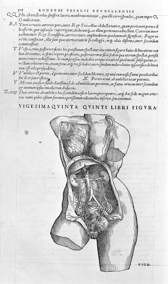
When Andreas Vesalius commissioned the famous illustrations of the interior of the body for his 1543 Fabrica, he was restricted to two dimensions, and to black and white. He covered both the outside and the inside, in particular in the whole-body images, where a male cadaver strides through the landscape around the city of Padua, gradually losing bits of his body as he goes. However, there are very few illustrations of the female body in Vesalius, as the male is used to show the general “human” body and the female is used only to show the organs of reproduction. In the smaller images illustrating the text, both the male and the female body are seen as if made from cold stone, using the “broken statue” style. One interpretation is that this is a deliberate reference to the classical past, part of Vesalius’s balance between respecting the past while, on some topics, challenging the long-established authorities, and the reuse in some illustrations of the ancient statue known as the Belvedere Torso – discovered in Rome in around 1490 – supports that idea. The missing parts also act to remove any sense of the original human model for each image; was there one person, or are these really an amalgam of many cadavers?[21] But disturbing hints of individuality sometimes remain; in the female body on the right of Figure 25 (Ill. 1) over the woman’s left shoulder appears what could be a piece of hair, or could perhaps hint at a rope from when the woman was hanged, because execution was the source of Vesalius’s bodies.
For the female body, perhaps the best-known illustration is Figure 27 (Ill. 2) which, as is often noted today, looks more like a penis than a womb. Most influentially, this was the claim of Thomas Laqueur, as part of his now-refuted model which held that, in pre-eighteenth-century Europe, the female body was seen as an outside-in version of the male body: he labels Vesalius’s Figure 27 “Vagina as penis” and states that “In this world the vagina is imagined as an interior penis” and “A whole world view makes the vagina look like a penis to Renaissance observers.”[22] But it is not the Renaissance readers who saw a penis here: it is us, the modern readers. Renaissance readers, and those who read their work in the early modern period, instead challenged the model: the English translation of the work of the Bartholins (father and son) included “The similitude of the yard [i.e. the penis] and of the womb, ridiculous.” “Ridiculous” here translates the Latin ineptus.[23] When he wrote “the womb,” he was including the vagina in this term. The vagina was not regarded as a separate part in the pre-modern world ; instead, following the terminology of the ancient world, it was “the neck of the womb,” uteri cervix. In Galen, this part stretches and dilates to provide “an unblocked road for the semen.”[24] Our cervix and our womb were not the equivalent of the pre-modern cervix and womb.
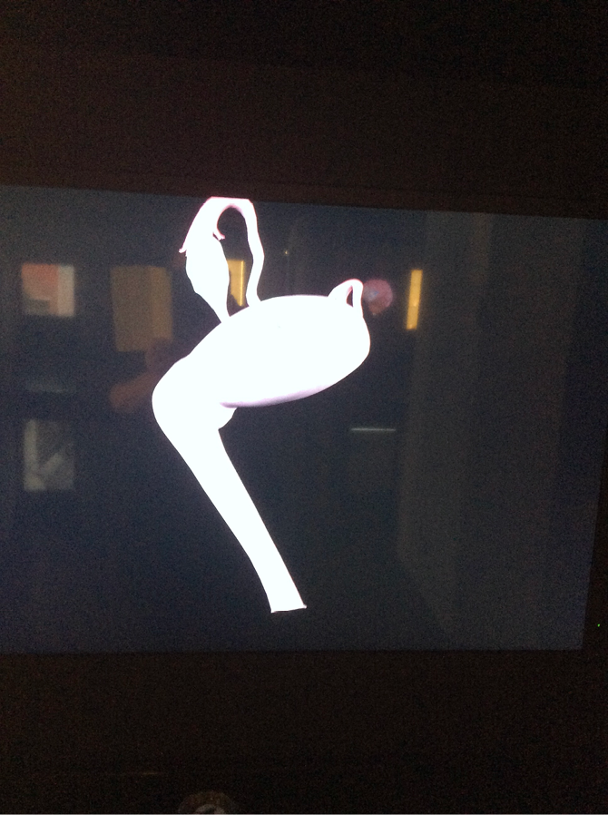
The features of the womb in Figure 27 reflect the fact that it has been cut in such a way that it is nothing like what we expect to see, by omitting the Fallopian tubes and ovaries. Today, these parts are familiar to us from numerous modern textbooks and websites, as in this image from a display at MUSME, the Museo di Storia della Medicina in Padua, which, unusually, also gives some sense of the relative angles of womb and vagina rather than “flattening” these (Ill. 3). Tubes and ovaries are included in most modern images because we regard all the reproductive organs as a single structure or system, but that is our decision; and, even when the tubes and their function were understood, wombs were often shown without them, because the main interest of those producing the illustrations lay in obstetrics, and in the implications for delivery of the baby’s position in utero.[25]
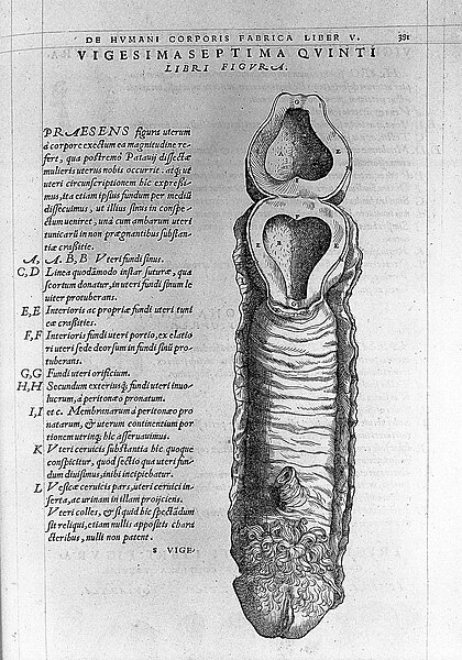
The dissection leading to Figure 27 has been performed not only to emphasise that the womb, vagina, and external genitalia form a single structure, but even more importantly to show that the cavity of the womb is empty. This is clear from the text, where Vesalius states that “This figure concerns, with respect to its size, the womb cut out of the body, in the manner of the womb of the last woman we dissected at Padua” and this is followed by a reference to the “thickness” of the tunics or layers of the womb here “in women who are not pregnant.”[26] That, then, is the real message of the image: that the woman whose womb is shown is not pregnant, and it has been cut open to show that there is nothing in it. This is important because it means that the woman from whom it was taken died according to the law, which stated that a pregnant woman could not be executed ; it was necessary to wait until she had given birth to the baby. Vesalius tells us that the woman whose womb is represented here also features in a second image, this time of her torso (Figure 24), and Katy Park has further identified her as the woman on the iconic title page.[27] This woman was middle-aged, had given birth many times and, having been condemned to be hanged for a crime that is nowhere identified, she had tried to avoid her sentence by claiming to be pregnant. Vesalius’s exposure of her womb on the title page image and in Figure 27 emphasises that women’s own words cannot be trusted as evidence of their pregnancy – or lack of it. However, as the midwives who had examined her had said she was not in fact pregnant, Vesalius’s findings also support their expertise. Here, then, reality is not being trimmed to fit a pre-existing Galenic theory, but the body is being used to answer a very specific query about whether or not the real woman whose womb is shown was pregnant. Perhaps, then, we should resist seeing it as a generic ‘womb’ image, and instead understand it as the specific womb of a specific woman.
But the range of materials of which images of the womb have been constructed is far greater than pen and ink; it expanded in the eighteenth century and continues to grow. Over the history of western medicine, wombs have been made in terracotta, wax, papier-mâché, wood, porcelain, metal, and rubber; since the mid-twentieth century, we can add wool, to which I shall return in my final section.
Wombs in three dimensions
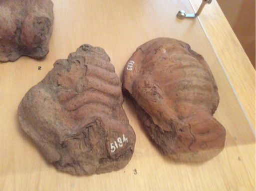
The earliest three-dimensional models of the womb may be votive offerings from the ancient Greek and Roman worlds.[28] Often labelled as “wombs” in museum catalogues and exhibitions, these are typically terracotta mould-made models showing a ribbed organ with a mouth[29] (Ill. 4). Some have extra chambers at the side: others have more complex ribbing or different decoration. Some contain small clay spheres which make a noise when the object is shaken or moved. There is increasing awareness that our identification of all of them as “wombs” may be misleading, and that the absence of human dissection in this period should also be taken into account; as Princeton Art Museum’s website points out, “This widespread interpretation does not explain why these corded objects do not really look like wombs, which most ancient people would never have seen.”[30] They could instead be any of the “container organs” of the body.
We have no evidence that these terracotta possible-wombs were painted, but other ancient votives do show traces of paint, so this is not impossible. Martin Söderlind has studied votive heads from central Italy, noting that we can estimate how many were made by looking at more and less blurry heads produced; maybe more than 200 being made during the life of one mould.[31] In these heads, the back was handmade while the front was not. Some had traces of paint; hair in a dark colour, red for male faces and white for female faces, red for the iris, black for the pupil.[32] These are mass-produced and mass-marketed objects, but have the potential for personalisation by the individual dedicant. Even if originally generic, once someone buys a body votive to present to the god or goddess, does it become somehow the womb of the person presenting it, or in whose name it is presented? We are told that visitors to healing sanctuaries in antiquity would wander around reading the inscriptions and looking at the objects, but we do not know what an ancient Greek woman would have thought of seeing wombs on display in a temple.[33]
Wax, a particularly powerful medium used for bodies and body parts since the early modern period, can not only be coloured to look lifelike, but also has the additional advantage – or, depending on its context, disadvantage – of appearing “wet.”[34] As Georges Didi-Hubermann noted, “there probably exists no other substance that can imitate with such polyvalence both the external flesh, the skin, and all the internal flesh, the muscles and the viscera.”[35] Surviving, although of course often restored, anatomical waxes such as Clemente Susini’s Anatomical Venus (c. 1790) from Florence are disturbing to viewers because of this fresh, wet appearance.[36] At what Joanna Ebenstein called “the last stage of anatomical striptease”, each of the anatomical Venus figures also turns out to contain a foetus.[37] That, after all, is – historically – the point of the female body.
The contrast between the apparent wetness of wax and the dead dryness of a modern body part from Gunther von Hagens’ Body Worlds exhibitions is striking. For the contemporary viewer of von Hagens’ work, which uses real human and animal bodies treated by his plastination process, seeing a real (plastinated) body can be experienced as less upsetting than an artistic creation in wax, because the wax looks more ‘wet’ and thus closer to life than the treated cadavers.[38] Modern work in wax, such as the artist John Isaacs’ A Necessary Change of Heart (2000), includes using the medium to show the human body, partially dissected, as if butchered. Unlike the bloodless historical wax models, this one deliberately suggests fresh blood.[39]
Terracotta and wax were by no means the only materials used to create three-dimensional wombs and other body parts. Some three-dimensional wombs were a feature of eighteenth-century obstetrical education, being used to demonstrate birth positions and difficult deliveries. The womb, rather than the woman, took centre stage in these models, as was also the case in the two-dimensional images of William Hunter’s Anatomy of the Human Gravid Uterus of 1774. In Hunter’s illustrations, if the woman was present at all, it was as the remains of a pair of amputated legs.[40] In Bologna, a glass womb was used to train both men and women practitioners; the trainee midwife or obstetrician could be blindfolded and try to work out from the sense of feel what was happening inside the womb, while the rest of the trainees watched.[41]
Another material was wood, as in a mixed-media French model by Pierre Constant Budin and Alphonse Pinard, first described in 1877, including a latex abdomen and a latex womb containing a dead foetus and some water. The more expensive models included a screw on the back which changed the curvature of the spinal column. Cloth on its own could also be used, although sometimes a real human pelvis formed the base.[42] The controversial element here was the baby: there was much debate about whether a cloth one was sufficient, or whether the best method was to use a preserved stillborn child.[43]
By the nineteenth century, rubber was also being used. The glass wombs of the eighteenth century could include fluids, used to give a sense of the fluids of birth, but rubber had even more potential. These could be used in ‘mannikins’ (also known as phantoms) where a complete woman was modelled, as in one illustrated in 1890, made in Germany and exhibited at the Obstetrical Society of Philadelphia in 1889.[44] We may find the presence of a face and of clothing very disturbing here, but the use of stockings is also something found in models used in the eighteenth century. One possible womb to accompany this mannikin was made of rubber with what is apparently also a rubber cervix, which could be tightened and loosened. The mannikin also included a rubber rectum. The pouch at the side of the womb could be filled with water, to give the impression of amniotic fluid. Another material used was ivory, perhaps with the blood vessels and cord rendered in red wool. Print, glass, wood, cloth, ivory, and rubber are all giving a similar message; the focus is on the baby and removing it successfully, rather than on the mother’s body.
Woollen wombs
The use of knitting in the twenty-first century seems to be something very different; the “knit your own body parts” movement changes the whole tone of what is being done, and how the womb is represented. In contrast to the complex representations of the womb in other media across history, knitting and stitching are accessible, democratic ways of making the material body visible. Hand-knitting has a long history, most commonly done as a source of income or as a way of saving money on clothes: we knit to keep warm. Working with fabric and thread, from weaving to embroidery, has in the West largely been understood as women’s work, and as part of a woman’s household tasks, although it was also a marketable skill.[45] There have been some changes over time, in western history, about how each of these forms of work has been gendered and valued: for example, in the medieval world, men and women worked together in embroidery workshops; in the Renaissance, most professional needleworkers were men[46], but by the eighteenth century embroidery was gendered as exclusively female, one of the ‘accomplishments’ an elite woman was supposed to demonstrate. Rozsika Parker’s The Subversive Stitch, first published in 1984, has not only been reprinted but has now been issued in a revised edition: she shows how embroidery was the proper occupation of a woman through Western European history, but also how it came to be re-used by women activists, such as the suffragettes, for their own purposes.[47]
The movement to encourage the knitting of body parts can be seen as a further politicization of working with thread and fabric. For many knitters, this is a hobby not an essential household task; no longer about saving money or about making wearable garments, it has become a leisure activity, although some makers are selling their work online. Yet knitting is far less “tame” than this may suggest; it is no longer utilitarian, done to keep warm, but can have entirely different purposes.
Knitted body parts often challenge the lines between art and craft, between public and personal. Rosina Godwin’s Knitted Womb (2017) was marketed as a piece of sculpture; the artist has described how here work is subverting “the nurturing associations of textiles.”[48] Such art objects are not my focus here; I am concentrating on ordinary women using knitting for their own purposes.
In contrast to some of the other media in which the womb has been created, in knitted wombs the focus is almost always on the external appearance. A key aspect is the properties of wool itself; as we have already seen, wax looks “wet,” thus persuasively and disturbingly lifelike, worryingly “real” for the viewer. Knitting – even of internal organs – is always going to look soft, friendly and accessible.[49] But there may be something even deeper in our psyche about wool, and women: in the Hippocratic medical texts, in terms of the texture of their flesh, woman is like raw wool, man is like the finished woven garment, and this is why women absorb more fluid and need to menstruate to release it.[50] There is an interesting challenge for the designer of body parts patterns in converting flesh into wool, wet into dry, and in finding the right colours and the right textures. While knitted wombs may be used for educational purposes, they are more likely to be used for pleasure. Some, however, are explicitly political in their purpose.
Knitted body parts with clear educational purposes include those made in 2016 by a team based at the University of Central Lancashire, who created them for local primary schools; the adult knitters have commented that their own knowledge of the body grew as a result of making these objects.[51] Knitted wombs, specifically, have been used in UK Parentcraft classes and by midwives educating pregnant women since the 1960s, when the National Childbirth Trust produced a pattern.[52] They are currently available with an added foetal doll and placenta, which “help[s] learners understand the sequence of events during labour, from the baby’s descent through the birth canal to the delivery of the placenta.”[53] The earliest use of something like this is in around 1940, in a teaching demonstration at the American College of Surgeons, reported in Time magazine for that year.[54] A balloon or beach ball is put into the knitted womb, with a smaller hard ball in the cuff; elastic can be threaded through the bottom and top of the cuff. If you palpate the large ball, the top of the small ball will appear “to represent dilatation of the cervix”. As the cervix dilates, it also becomes thinner. This is something which could not be seen by learners in any other way, as it is happening inside the body, invisible to them.[55] Here, the properties of wool are a particularly appropriate way of imitating the properties of the body.
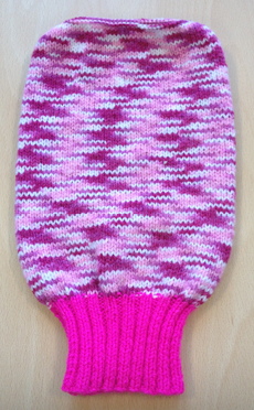
The instructions on the National Childbirth Trust pattern for knitting a womb include: “No attempt should be made to give this visual aid a lifelike appearance; it will probably be more acceptable to classes if knitted in plain colours – possibly buff or oatmeal, not bright colours.” These instructions were ignored to excellent effect by Sue Tully, a midwifery lecturer at Bournemouth University, who used pink (Ill. 5). But this is not as extreme a variation as the one described in an article in The Guardian, where in 2013 the journalist Suzanne Moore described her experience of attending ante-natal classes; the midwife taking these
had knitted a uterus. She apologised for the fact that it was navy blue as she had run out of pink wool. The plain knit was the uterus and the ribbed sock bit was the cervix. She then proceeded to push a tennis ball through it. That, she said, was “birth.”[56]
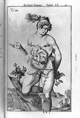
The “navy blue” uterus here recalls a series of images using, not a ball, but a Cabbage Patch Kid doll; these cloth dolls with plastic heads were first made in 1982, and at one point a series of images combining one of these dolls with a knitted blue womb could be seen online.[57] Videos have been posted in which viewers can “Watch a Cabbage Patch Doll Being Born.”[58] Colin Bailey has pointed out that the cabbage was a symbol for the womb in sixteenth-century Europe, linking this to the idea that babies appear in the cabbage patch, as in the French “venir au monde sous un chou,” which dates to the eighteenth century.[59] In a famous image of the womb from Spigelius’s De formato foetu, the baby does indeed appear to be nestling in a cabbage[60] (Ill. 6).
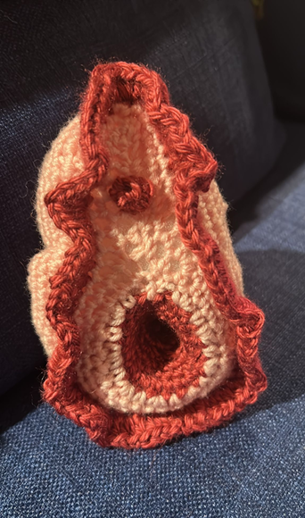
Knitted wombs are still marketed as teaching aids and may include ovaries, Fallopian tubes, ligaments, and blood vessels as well as a placenta.[61] These are very practical wombs. But today the main use of knitting and crochet is not by professionals, but by ordinary women: for ornamental purposes, sometimes for therapeutic purposes, but often to challenge negative messages about the female body, and to empower women to be confident about their wombs and about other parts of their bodies (Ill. 7).
Knitting can be about community: people often knit together. Creating knitted body parts is made much easier by the internet, which connects people internationally; websites for knitters not only share patterns but also discuss at great length these questions of colour and texture, and what is the best wool to use. The website Stitchin’ Bints published a roundup of body parts patterns in 2013.[62] Knitted or crochet body parts for which patterns are widely available online include gifts to cheer up a woman who has had surgery: for example, a hysterectomy, or the removal of an ovarian cyst (one such knitted womb was named “Ursula Uterus”).[63] I wonder whether there is also some vestige here of the terracotta votive womb, originally given to the gods to request or to give thanks for healing ? Canadian feminist Beryl Tsang’s “tit bits” were created after her mastectomy.[64] She wanted to find a breast prosthesis but thought the one she had bought looked like “raw liver.” Instead, she decided to knit her own and wear it with one of her favourite bras. She encourages other women to knit a personal prosthesis; this means women are not dependent on the commercial medical market.[65]
All patterns for knitted wombs made for pleasure rather than for education seem to go back to the first body parts pattern offered by knitty.com, in winter 2004, created by the knitter and crocheter MK Carroll, who is based in Hawai’i. The designer noted of this womb that “It’s not completely anatomically accurate. I’ve taken a few liberties with the general shape and scale, as well as leaving out the ligaments connected to the ovaries. And, of course, the human uterus is not normally bubblegum pink.”[66] Pink is of course the typically feminine colour, and its use in knitting is often consciously ironic. As noted by Beth Ann Pentney in a 2008 article asking “Are the fibre arts a viable model for feminist political action?”, books on knitting can now have an “ironic tone and kitschy visuals.”[67]
The instructions for Carroll’s womb include “Stuff body until it feels firm but cuddly.” “Cuddly” is not a word one would often use in connection with the womb. Omitting the ligaments makes this womb more like a Hippocratic one, free to wander around the body. This wandering womb model has been a powerful one in the reception of the ancient body in medicine from the medieval world to today, not least because it is so alien to us now, and Carroll refers in her own work to such popular understandings of the classical medical tradition. She wrote in 2007: “Hippocrates coined the term hysteria, and thought that it was caused by the womb traveling around the body in search of children. This womb will be doing her traveling outside of my body.”[68] Carroll’s womb is not commercial; she insists that wombs made from her pattern cannot be sold, and only the costs of materials can be met. If you want to add ovaries, she suggests “going with the ovaries your Womb prefers – if she wants Swarovski crystals, give them to her. Pearls ? Nothing is too good for your womb.”[69]
As these extracts from Carroll suggest, the knitted womb can bring a sense of fun, even of naughtiness. I somehow doubt that an ancient Roman terracotta womb was ever “fun” in this way. The knitted womb can be seen as part of what was described in 2012 as a “new frankness” in discussing the female body.[70] In the words of Amy Singer on knitty.blog, “Why a knitted womb ? Because... (a) it’s cute (b) we can (c) it’s really freaking cute, and who would have ever thought you could knit a CUTE WOMB?”[71]
But, as Carroll’s decision to omit the ligaments demonstrates, choices remain in how you create a “cute womb.” Zabet Stewart’s “Snatchel” makes the womb into a bag, focusing not on Carroll’s animate and travelling womb, but instead on the aspect of the womb as container. She has sparkling ovaries, and a drawstring which perhaps takes the place of the ligaments. Her womb not only includes the ovaries, but also has more sense of the vagina, and includes the labia and pubic hair. She noted that she “can’t take credit for the clever name, however, that was Erik, from our Stitch ‘n Bitch. I had called it something uninspired and utilitarian like Vagina Bag or Vulva Purse. Thank the Universe for Erik. There’s nothing worse than a utilitarian vulva. She may not be pretty, but she’s mine.”[72] The instructions for the snatchel include:
Now, Labia Minora vary greatly from woman to woman. I did a single round of single crochet and randomly placed 1 or 2 chain sts in between the single crochets as I went around. This gave my Labia Minora a ruffled, irregular look. If you want your Labia Minora to protrude past the Labia Majora, as women’s do, you might want to try 2 or 3 rounds of single crochet. If this is the case, I recommend only adding random extra chain stitches on the top row.
The amazement at a “cute womb,” picked up in Stewart’s “she may not be pretty, but she’s mine,” is at least partly a reaction against contemporary women’s unease about the appearance of their body parts; knitters are reclaiming the womb as something to be celebrated.
While knitted and crochet female body parts started off as fun, as “because we can,” and as educational, they are now increasingly politicised, even beyond the politics of the body which offering a body positive approach can bring. They are part of what has been called “craftivism,” the use of craft for political or social causes.[73] This includes “radical knitting,” where objects such as army tanks are covered in pink crochet squares. Again, pink: to feminise them. An American campaign in 2005, “Knit4Choice” or “Wombs on Washington”, aimed to leave knitted wombs at the Supreme Court in Washington DC to protest against threats to restrict access to abortion.[74] However, this never took place, not least because of divisions in the organising group. In the US 2012 Government Free V-JJ campaign, “Knit your congressman a vagina” (V-JJ is an abbreviation for “vagina”)[75], the slogan was “If they have their own, they can leave ours alone!” Under “Who Are We?” the movement states “We are women, we are strong, we are smart. And we have a sense of humor.” The 2011-2013 felt cervix project attempted both to encourage awareness of cancer of the cervix, and to encourage women to vote for political candidates committed to women’s reproductive rights.[76]
The combination of humour, advocacy for a body positive approach, and a political message is also found in a patchwork “vulva quilt” made by the UK’s Shoreditch Sisters, a “modern WI” or Women’s Institute, one of a series of local groups more traditionally associated with making jam, but which have long practised a form of “stealth feminism.”[77] The Shoreditch Sisters linked this project to raising awareness of female genital mutilation and attacking the cult of the “designer vagina,” as well as rejecting “the stigma and disgust people have for our natural organs.”[78] After a day of making these at the Women’s Library they blogged “I have honestly never said the word vagina so many times in one day, only to be reminded afterwards that the vagina is technically the inside bit and we were making vulvas, the exterior bit.”[79] This is an interesting comment, raising the issues of what the right words may be, as well as which ones can be spoken aloud.
Like the creation of the vulva quilt, Stewart’s Stitch n’ Bitch group, and indeed the Shoreditch Sisters’ Knit and Natter group, the Whitstable Profanity Embroidery Group is partly about building community for women.[80] Founded in 2014, it took its inspiration from an image of an elderly lady embroidering “Fuck the World” on a cushion.[81] They meet every month in a local pub, where they “drink, stitch and chat.” No prior skills are required; in Pentney’s terms, this is “creativity in spite of artistic skill or training.” As well as embroidering swear words, they work with slang terms for parts of the female body. In 2019 they were featured in a TV series, “All Women,” where they were described as a punk version of the WI.[82] A former colleague of mine, Kim Shahabudin, sells simple, subversive cross-stitch kits of single words with sexual connotations, marketed as “Smut and tut” (“smut” being something indecent or obscene, and “tut, tut” being what one says to express disapproval).[83] The Vagina Museum in London has also sold patterns for knitting or crocheting various body parts, among them clitorises.[84]
The history of women as producers of fabric not only goes back a very long way, but also comes with an awareness of women’s craftiness, in both senses of the word: both their role as producers of craft work, but also the cunning to which their structural position in society may make them resort. Dora Wheeler’s silk piece, “Penelope unravelling her work at night” (1886) famously presents Penelope both as a model of fidelity – loyal to her absent husband Odysseus – and of deviousness: she has sworn to choose one of the suitors to marry but will only do this when the shroud she is weaving is completed.[85] Beth Ann Pentney commented: “If second-wave feminists have been historicized as women who put down their knitting, third-wave feminists may be characterized as those who have picked it back up again.” When they pick up their knitting, they are being crafty in many ways ; as makers, and as women undercutting the messages about their organs of generation which centuries of medical writing have created.
Over the history of the body in western Europe, many different materials have been used to represent the womb, all of them carrying more meaning than simply an attempt at realistic portrayal of its appearance or powers. These materials include words, which can reflect how the womb is understood and valued, whether as “housewife” or as “sewer.” But they also include different physical media, some of them three-dimensional. Wool can give a message of accessibility, a long way from the operating theatre or the cadaver. Textbook images of what we “should” look like appear cold and unfriendly, whereas wool is approachable: wool is “cute.” Emily Stoneking’s aKNITomy project involves knitted animal dissections ; she works at “the places where art and science intersect” and comments “I am also interested in using cuddly materials (like cozy knitting) to create objects that many people are usually squeamish about.”[86] The knitted wombs draw on a similar sense of reducing anxiety. The knitted womb is neither a miraculous organ, nor a sewer. It is playful, fun, and it knows what it wants: a positive approach to the body, and reproductive rights. Yet, at the same time as knitting reduces the power of the womb to shock when it is performed within a women’s group, knitted wombs can also be used precisely to disturb the male viewer, as when women use them to persuade legislators not to limit women’s reproductive rights.
Auteurs
Bibliography
Anon., Anathomia oder abconterfectung eines weybs leyb: wie er innwendig gestaltet ist, Strassburg, Heinrich Vogtherr, 1538
Azzolini, Monica, “Exploring Generation: A Context to Leonardo’s Anatomies of the Female and Male Body”, in Leonardo da Vinci’s Anatomical World: Language, Context and “Disegno”, ed. Alessandro Nova, Domenico Laurenza, Florence, Marsilio, 2011, p. 79-97
Bailey, Colin B., “‘Details that surreptitiously explain’: Boucher as a Genre Painter”, in Rethinking Boucher: Issues and Debates, ed. Melissa Hyde, Mark Ledbury, Los Angeles, Getty Research Institute, 2006, p. 39-60
Bartholin, Caspar, Anatomicae institutiones corporis humanis, [Wittenburg], A. Rüdinger, 1611
Bartholin, Thomas, Bartholinus Anatomy; made from the precepts of his father, and from the observations of all modern anatomists; together with his own ... in four books and four manuals ... / published by Nich. Culpeper and Abdiah Cole, London, Peter Cole, 1663
Bottoni, Albertino, De morbis muliebribus, in Gynaeciorum, ed. Caspar Bauhin, Basle, Thomas Guarinus, 1586-1588
Bradley, Mark, Victoria Leonard, Laurence Totelin, ed., Bodily Fluids in Antiquity, London, Routledge, 2021, https://doi.org/10.4324/9780429438974
Connor, J. T. H., “‘Faux Reality’ Show? The Body Worlds Phenomenon and its Reinvention of Anatomical Spectacle,” Bulletin of the History of Medicine, 2007, t. 81, no 4, p. 848-862, https://doi.org/10.1353/bhm.2007.0112
Crooke, Helkiah, Microcosmographia: A Description of the Body of Man, London, William Jaggard, 1615
Crowther, Kathleen M., Adam and Eve in the Protestant Reformation, Cambridge, Cambridge University Press, 2010
Da Monte, Giambattista, De uterinis affectibus, Venice, B. Constantinus, 1554
Dacome, Lucia, “A Crystal Womb”, in Reproduction: Antiquity to the Present Day, ed. Nick Hopwood, Rebecca Flemming, Lauren Kassell, Cambridge, Cambridge University Press, 2018, https://doi.org/10.1017/9781107705647.073
Dacome, Lucia, “Waxworks and the Performance of Anatomy in Mid-18th Century Italy”, Endeavour, 2006, t. 30, no 1, p. 29-35, https://doi.org/10.1016/j.endeavour.2006.01.004
Dacome, Lucia, “Women, Wax, and Anatomy in the ‘Century of Things’”, Renaissance Studies, 2007, t. 21, no 4, p. 522-550, https://doi.org/10.1111/j.1477-4658.2007.00461.x
Didi-Huberman, Georges, “Wax Flesh, Vicious Circles”, in Encyclopaedia Anatomica: A Complete Collection of Anatomical Waxes, ed. Monika von Düring, Georges Didi-Huberman, Marta Poggesi, Cologne, Taschen, 1999, p. 64-74
Dillon, Matthew P. J., “The didactic nature of the Epidaurian iamata,” Zeitschrift für Papyrologie und Epigraphik, 1994, t. 101, p. 239-260
Dillon, Matthew P. J., Pilgrims and Pilgrimage in Ancient Greece, New York, Routledge, 1997
Dionis, Pierre, A General Treatise of Midwifery, London, A. Bell et al., 1719
Donaldson, Iain M. K., “Smellie & Hunter: Atlases of the Gravid Uterus. Part 2,” Journal of the Royal College of Physicians, Edinburgh, 2016, t. 46, p. 140-142, https://doi.org/10.4997/jrcpe.2016.115
Dougal, Daniel, “The Teaching of Practical Obstetrics,” British Journal of Obstetrics and Gynaecology of the British Empire, 1933, t. 40, p. 99-102, https://doi.org/10.1111/j.1471-0528.1933.tb15525.x
Du Laurens, André, Historia anatomica humani corporis, Heidelbergae, Johannes Rhodius, 1602
Dubois, Jacques, Livre de la nature et utilité des moys des femmes, Paris, Morel, 1559
Ebenstein, Joanna, “Ode to an Anatomical Venus,” Women’s Studies Quarterly, 2012, t. 40, no 3/4, p. 346-352, https://doi.org/10.1353/wsq.2013.0021
Edgar, James Clifton, “The Manikin in Teaching Obstetrics,” New York Medical Journal, 1890, t. 52.
Falloppio, Gabriele, Observationes anatomicae, Marco Antonio Ulmo e Gratioso Perchachino, Venice, 1561
Fissell, Mary, Vernacular Bodies: The Politics of Reproduction in Early Modern England, Oxford, Oxford University Press, 2004, https://doi.org/10.1093/oso/9780199269884.001.0001
Flemming, Rebecca, “Wombs for the Gods”, in Bodies of Evidence: Ancient Anatomical Votives Past, Present and Future, ed. Jane Draycott, Emma-Jayne Graham, London, Routledge, 2017, p.112-130, https://doi.org/10.4324/9781315096193-7
Green, Monica, The Trotula: A Medieval Compendium of Woman’s Medicine, Philadelphia, University of Philadelphia Press, 2001, https://doi.org/10.9783/9780812204698
Guillemeau, Jacques, Childe-Birth, or the Happy Deliverie of Women, London, A. Hatfield, 1612
Harcourt, Glenn, “Andreas Vesalius and the Anatomy of Antique Sculpture,” Representations, 1987, t. 17, p. 28-61, https://doi.org/10.2307/3043792
Harrison, Katherine, Ogden, Cassandra A., “Knit ‘n’ Natter: A Feminist Methodological Assessment of Using Creative ‘Women’s Work’ in Focus Groups”, Qualitative Research, 2020, p. 1-17, https://doi.org/10.1177/1468794120945133
Herrlinger, Robert, Feiner, Edith, “Why did Vesalius Not Discover the Fallopian Tubes?” Medical History, 1964, t. 8, no 4, p. 335-341, https://doi.org/10.1017/S002572730002980X
Jordanova, Ludmilla, Nature Displayed: Gender, Science and Medicine 1760-1820, New York, Addison Wesley Longman Inc, 1999
Keller, Eve, Generating Bodies and Gendered Selves: The Rhetoric of Reproduction in Early Modern England, Seattle, University of Washington Press, 2007
Kemp, Martin, Wallace, Marina, Catalogue, Spectacular Bodies: The Art and Science of the Human Body from Leonardo to Now, Berkeley, LA and London, University of California Press/Hayward Gallery, 2000
King, Helen, Hippocrates’ Woman: Reading the Female Body in Ancient Greece, London, Routledge, 1998
King, Helen, Immaculate Forms: Uncovering the History of Women’s Bodies, London, Profile, 2024
King, Helen, “Inside and Outside, Cavities and Containers: The Organs of Generation in Seventeenth-Century English Medicine” in Medicine and Space: Body, Surroundings and Borders in Antiquity and the Middle Ages, ed. Patricia A. Baker, Han Nijdam, Karine van ‘t Land, Visualising the Middle Ages (4), Leiden, Brill, 2011, p. 37-60, https://doi.org/10.1163/9789004226500_004
King, Helen, Midwifery, Obstetrics and the Rise of Gynaecology: The Uses of a Sixteenth-century Compendium, Aldershot, Ashgate, 2007
King, Helen, The One-Sex Body on Trial: The Classical and Early Modern Evidence, Farnham, Ashgate, 2013
Kusukawa, Sachiko, Picturing the Book of Nature: Image, Text, and Argument in Sixteenth-Century Human Anatomy and Medical Botany, Chicago, University of Chicago Press, 2012, https://doi.org/10.7208/chicago/9780226465289.001.0001
Laqueur, Thomas, Making Sex: Body and Gender from the Greeks to Freud, Cambridge, MA and London, Harvard University Press, 1990
Lemay, Helen Rodnite, Women’s Secrets: A Translation of Pseudo-Albertus Magnus’ De secretis mulierum with Commentaries, Albany, State University of New York Press, 1992
Nott, John, Harris, Anita, “Sticky Models: History as Friction in Obstetric Education”, Medical Anthropology Theory, 2020, t. 7, no 1, p. 44-65, https://doi.org/10.17157/mat.7.1.738
Owen, Harry, Pelosi, Marco A., “A Historical Examination of the Budin-Pinard Phantom: What Can Contemporary Obstetrics Education Learn from Simulators of the Past?,” Academic Medicine: Journal of the Association of American Medical Colleges, 2013, t. 88, no 5, p. 652-656, https://doi.org/10.1097/ACM.0b013e31828b0464
Park, Katharine, Secrets of Women: Gender, Generation, and the Origins of Human Dissection, New York, Zone Books, 2006
Parker, Rozsika, Pollock, Griselda, Old Mistresses: Women, Art and Ideology, London, Routledge & Kegan Paul, 1981
Parker, Rozsika, The Subversive Stitch: Embroidery and the Making of the Feminine, London, The Women’s Press, 1984
Paster, Gail Kern, The Body Embarrassed: Drama and the Disciplines of Shame in Early Modern England, Ithaca, NY, Cornell University Press, 1993, https://doi.org/10.7591/9781501724497
Pentney, Beth Ann, “Knitting and Feminism’s Third Wave: Are the Fibre Arts a Viable Model for Feminist Political Action?,” thirdspace: a journal of feminist theory and culture, 2008, t. 8, no 1
Read, Sara, Menstruation and the Female Body in Early Modern England, London, Palgrave, 2013, https://doi.org/10.1057/9781137355034
Richardson, William, Carman, John, On the Fabric of the Human Body: A Translation of De Humana Corporis Fabrica Libri Septem. Book V, Novato, CA, Norman Publishing, 2007
Searle, Karen, Knitting Art: 150 Innovative Works from 18 Contemporary Artists, Beverly, MA, Quarto Press, 2008
Siraisi, Nancy, “Vesalius and the Reading of Galen’s Teleology,” Renaissance Quarterly, 1997, t. 50, p. 14-28, https://doi.org/10.2307/3039327
Smith, Chloe Wigston, Women, Work, and Clothes in the Eighteenth-Century Novel, Cambridge, Cambridge University Press, 2013, https://doi.org/10.1017/CBO9781139542708
Söderlind, Martin, Late Etruscan Votive Heads from Tessennano: Production, Distribution, Sociohistorical Context, Rome, L’Erma di Bretschneider, 2002
Spigelius, Adrianus, Opera quae extant omnia, Amsterdam, Johannes Blaue, 1645
Stolberg, Michael, “A Woman Down to her Bones. The Anatomy of Sexual Difference in the Sixteenth and Early Seventeenth Centuries,” Isis, 2003, t. 94, p. 274-299, https://doi.org/10.1086/379387
Turney, Joanne, The Culture of Knitting, Oxford, Berg Publishers, 2009
Voss, Heinz-Jürgen, Making Sex Revisited. Dekonstruktion des Geschlechts aus biologisch-medizinischer Perspektive, Bielefeld, transcript, 2010, https://doi.org/10.1515/9783839413296
Walter, Tony, “Body Worlds: Clinical Detachment and Anatomical Awe,” Sociology of Health and Illness, 2004, t. 26, no 4, p. 464-488, https://doi.org/10.1111/j.0141-9889.2004.00401.x
Wills, Kerry, The Close-knit Circle: American Knitters Today, Westport, CT, Praeger Publishers, 2007, https://doi.org/10.5040/9798400627682
Notes
- 1 G. Kern Paster, The Body Embarrassed: Drama and the Disciplines of Shame in Early Modern England, Ithaca, NY, Cornell University Press, 1993, p. 2-4; for the ancient Mediterranean, see now the collection edited by M. Bradley, V. Leonard and L. Totelin, Bodily Fluids in Antiquity, London, Routledge, 2021; H. King, “Inside and Outside, Cavities and Containers: The Organs of Generation in Seventeenth-Century English Medicine”, in Medicine and Space: Body, Surroundings and Borders in Antiquity and the Middle Ages; Visualising the Middle Ages (4), ed. P. A. Baker, H. Nijdam, K. van ‘t Land, Leiden, Brill, 2011, p. 37-60.
- 2 G. Kern Paster, op. cit., p. 4, 7.
- 3 M. Green, The Trotula: A Medieval Compendium of Woman’s Medicine, Philadelphia, University of Philadelphia Press, 2001, p. 20, on how “the waste material was almost always thought to be collecting whether it issued from the body or not.”
- 4 G. Kern Paster, op. cit., p. 39. See also 24-25 on “the weaker vessel as leaky vessel.” On the Hippocratic treatises, H. King, Hippocrates’ Woman: Reading the Female Body in Ancient Greece, London, Routledge, 1998, p. 28-29.
- 5 M. Azzolini, “Exploring Generation: A Context to Leonardo’s Anatomies of the Female and Male Body”, in Leonardo da Vinci’s Anatomical World: Language, Context and “Disegno”, ed. A. Nova, D. Laurenza, Florence, Marsilio, 2011, p. 79-97. Some of the ideas in this section are developed in H. King, Immaculate Forms: Uncovering the History of Women’s Bodies, London, Profile, 2024, chap. 4.
- 6 Microcosmographia: A Description of the Body of Man, London, William Jaggard, 1615, p. 262-263; E. Keller, Generating Bodies and Gendered Selves: The Rhetoric of Reproduction in Early Modern England, Seattle, University of Washington Press, 2007, p. 68-69.
- 7 A. du Laurens, Historia anatomica humani corporis, Heidelbergae, Johannes Rhodius, 1602, p. 542 and Quaestio XI, p. 558-562, where he starts from the Hippocratic Places in Man 47 and its statement that the womb is the cause of all diseases of women.
- 8 H. R. Lemay, Women’s Secrets: A Translation of Pseudo-Albertus Magnus’ De secretis mulierum with Commentaries, Albany, State University of New York Press, 1992, p. 179 n. 119 observes that, despite him being referenced in this text, Avicenna is not the source of this image.
- 9 Th. Bartholin, Bartholinus Anatomy; made from the precepts of his father, and from the observations of all modern anatomists; together with his own ... in four books and four manuals ... / published by Nich. Culpeper and Abdiah Cole, London, Peter Cole, 1663, p. 70.
- 10 J. Oliver, Present for Teeming Women quoted in Sara Read, Menstruation and the Female Body in Early Modern England, London, Palgrave, 2013, p. 20.
- 11 P. Dionis, A General Treatise of Midwifery, London, A. Bell et al., 1719, p. 59. “Sewer” here translates the French « l’égout ».
- 12 Anathomia oder abconterfectung eines weybs leyb: wie er innwendig gestaltet ist, Strassburg, Heinrich Vogtherr, 1538, quoted in Kathleen M. Crowther, Adam and Eve in the Protestant Reformation, Cambridge, Cambridge University Press, 2010, p. 160-166.
- 13 G. da Monte, De uterinis affectibus, Venice, B. Constantinus, 1554.
- 14 Translations by Thomas Linacre and Niccoló Leoniceno; see H. King, Midwifery, Obstetrics and the Rise of Gynaecology: The Uses of a Sixteenth-century Compendium, Aldershot, Ashgate 2007, p. 53.
- 15 Th. Bartholin, op. cit., p. 65. Also in H. Crooke, op. cit., p. 223 and J. Guillemeau, Childe-Birth, or the Happy Deliverie of Women, London, A. Hatfield, 1612, p. 113; as all these early modern midwifery texts were dependent on each other, it was probably simply copied between them. Inter urinas et faeces nascimur is a phrase attributed to S. Augustine but there is much debate about its actual origin; see for example URL: https://artandpopularculture.com/Inter_faeces_et_urinam_nascimur, consulted on 19.02.2025.
- 16 A. Bottoni, De morbis muliebribus, in Gynaeciorum, ed. C. Bauhin, Basle, Thomas Guarinus, 1586-1588, vol. 2, ch. 7.
- 17 J. Dubois, Livre de la nature et utilité des moys des femmes, Paris, Morel, 1559, p. 120-121: the blood is « tres-utile ».
- 18 [Hippocrates], Epidemics 7.123; compare ta kata physin, “the natural things,” Epidemics 6.8.32, and Diseases of Women 3.230. On the significance of “Nature” in Vesalius, see N. Siraisi, “Vesalius and the Reading of Galen’s Teleology,” Renaissance Quarterly, 1997, t. 50, p. 14-28.
- 19 H. King, Midwifery, op. cit., p. 52.
- 20 Contra M. Fissell, Vernacular Bodies: The Politics of Reproduction in Early Modern England, Oxford, Oxford University Press, 2004, p. 60 which tries to make negative view of the womb into something novel, attributing it to the 1603 work of Edward Jorden: “Once the womb had been a wonderful body part, capable of transforming male and female seed into a new being, and marvellous in its ability to grow from the size of a walnut into a vessel containing a full-term baby. This positive view of the womb was challenged by a much darker vision, propounded in vernacular texts from 1603.” She tries to link this to the Reformation and women no longer thinking of themselves in terms of a pregnant Virgin Mary. Yet on the previous page she had noted – correctly – that “both positive and negative views of women’s bodies could be found in ancient learning” (p. 59). In fact, the features of Jorden’s references to the womb which she singles out as “startling” are all elements which go back to Galen.
- 21 G. Harcourt, “Andreas Vesalius and the Anatomy of Antique Sculpture,” Representations, 1987, t. 17, p. 28-61. The Belvedere Torso was acquired by Pope Julius II soon after its discovery and was on display in the Vatican gardens by the 1530s; on the various drawings of it, see S. Kusukawa, Picturing the Book of Nature: Image, Text, and Argument in Sixteenth-Century Human Anatomy and Medical Botany, Chicago, University of Chicago Press, 2012, p. 215.
- 22 Th. Laqueur, Making Sex: Body and Gender from the Greeks to Freud, Cambridge, MA and London, Harvard University Press, 1990, p. 4 (caption to Fig. 20); p. 82 also p. 313, the index entry to “Vagina as penis”. For challenges to Laqueur see M. Stolberg, “A Woman Down to her Bones. The Anatomy of Sexual Difference in the Sixteenth and Early Seventeenth Centuries,” Isis, 2003, t. 94, p. 274-299; H.-J. Voss, Making Sex Revisited. Dekonstruktion des Geschlechts aus biologisch-medizinischer Perspektive, Bielefeld, transcript, 2010; H. King, The One-Sex Body on Trial: The Classical and Early Modern Evidence, Farnham, Ashgate, 2013.
- 23 Th. Bartholin, op. cit., p. 62; C. Bartholin, Anatomicae institutiones corporis humanis, [Wittenburg], A. Rüdinger, 1611, p.114, on which see H. King, One-Sex Body, op. cit., p. 67.
- 24 Galen, Usefulness of Parts, 14.3, Kühn 4.148.
- 25 The Fabrica was of course published before any of this was known. The tubes were “discovered” by Vesalius’ pupil Gabriele Falloppio only in 1561 (he called them uteri tuba), so in Vesalius even their points of entry into the womb do not appear. See G. Falloppio, Obseruationes anatomicae, Venice, Marco Antonio Ulmo e Gratioso Perchachino, Venice 1561, p. 196vo on the uteri tuba and R. Herrlinger, E. Feiner, “Why did Vesalius Not Discover the Fallopian Tubes ?” Medical History, 1964, t. 8, no 4, p. 335-341.
- 26 A. Vesalius, Fabrica, op. cit., 1543, p. 381 (this should be numbered 481, but the pagination is incorrect from 313-391). Lat. “Praesens figura uterum a corpora exectum ea magnitudine refert, qua postremo Patauii dissectae mulieris uterus nobis occurrit” (the translation of W. Richardson, J. Carman, On the Fabric of the Human Body: A Translation of De Humana Corporis Fabrica Libri Septem. Book V, Novato, CA, Norman Publishing, 2007, p. 44: “This figure shows a uterus cut away from the body: it is the same size as the uterus of the woman who was the subject of our last dissection at Padua” misses Vesalius’ emphasis on size as the main purpose of the illustration). Vesalius goes on to explain that he has shown the womb opened so that the thickness or density of the cavity and tunics of the womb in a non-pregnant woman can be clearly seen; sinus ... una cum ambarum uteri tunicarum in non praegnantibus substantiae crassitie. On p. 533, Vesalius again refers specifically to Fig. 27 and notes – as in the caption – that this is the size of the womb in women who are not pregnant: ‘Vigesima septima tamen praesentis libri figura, eam uteri magnitudinem exprimit, quae frequenter in non praegnantibus, et praecipue in huius anni consectione Patauii occurrit’; the translation of Richardson, op. cit., p. 177 changes the emphasis on size and adds “often seen”, thus implying more about eye-witness evidence than does the Latin. I would translate as “For all that, Figure 27 of the present volume shows the size of the womb, as happens often in women who are not pregnant, and especially in a dissection at Padua in this very year”.
- 27 K. Park, Secrets of Women: Gender, Generation, and the Origins of Human Dissection, New York, Zone Books, 2006, p. 207 and p. 211. She was middle-aged, not – as Laqueur, p. 82 had earlier claimed – young.
- 28 R. Flemming, “Wombs for the Gods”, in Bodies of Evidence: Ancient Anatomical Votives Past, Present and Future, ed. J. Draycott, E.-J. Graham, London, Routledge, 2017, p. 112-130.
- 29 URL: https://wellcomecollection.org/works/tfpbygww, consulted 19.02.2025. The artist Becky Brewis has created gold votive wombs: URL: https://beckybrewis.com/Three-votive-wombs, consulted 19.02.2025.
- 30 URL: https://artmuseum.princeton.edu/collections/objects/5840, consulted 19.02.2025.
- 31 Late Etruscan Votive Heads from Tessennano: Production, Distribution, Sociohistorical Context, Rome, L’Erma di Bretschneider, 2002, p. 43.
- 32 M. Söderlind, op. cit., p. 266-267.
- 33 M. P. J. Dillon, “The didactic nature of the Epidaurian iamata,” Zeitschrift für Papyrologie und Epigraphik, 1994, t. 101, p. 239-260; M. P. J. Dillon, Pilgrims and Pilgrimage in Ancient Greece, New York, Routledge, 1997.
- 34 The wet/dry distinction is discussed in terms of clinical detachment by T. Walter, “Body Worlds: Clinical Detachment and Anatomical Awe,” Sociology of Health and Illness, 2004, t. 26, no 4, p. 464-488 and by J. T. H. Connor, “‘Faux Reality’ Show? The Body Worlds Phenomenon and its Reinvention of Anatomical Spectacle,” Bulletin of the History of Medicine, 2007, t. 81, no 4, p. 848-862.
- 35 G. Didi-Huberman, “Wax Flesh, Vicious Circles”, in Encyclopaedia Anatomica: A Complete Collection of Anatomical Waxes, ed. M. von Düring, G. Didi-Huberman, M. Poggesi, Cologne, Taschen, 1999, p. 64-74: quote on p. 65-66.
- 36 L. Dacome, “Waxworks and the Performance of Anatomy in Mid-18th Century Italy,” Endeavour, 2006, t. 30, no 1, p. 29-35; L. Dacome, “Women, Wax, and Anatomy in the ‘Century of Things’”, Renaissance Studies 2007, t. 21, no 4, p. 522-550.
- 37 J. Ebenstein, “Ode to an Anatomical Venus,” Women’s Studies Quarterly, 2012, t. 40, no 3/4, p. 346-352.
- 38 On von Hagens, see URL: https://bodyworlds.com/plastination/gunther-von-hagens/, consulted 19.02.2025; T. Walter, “Plastination for Display: A New Way to Dispose of the Dead,” Journal of the Royal Anthropological Institute, 2004, t. 10, p. 603-604, distinguishes between wet/dry and stable/unstable; a corpse is wet and unstable, while von Hagens’s plastinates are dry and stable.
- 39 M. Kemp, M. Wallace, Catalogue, Spectacular Bodies: The Art and Science of the Human Body from Leonardo to Now, Berkeley, LA and London, University of California Press/Hayward Gallery, 2000, p. 164-165: Isaacs’ work is illustrated on URL: https://www.kw-berlin.de/en/john-isaacs-a-necessary-change-of-heart/, consulted 19.02.2025. If I may share a personal reaction, when I saw this exhibition, this was the only object which made me feel physically sick, and I saw one other visitor rush out of the room where it was displayed.
- 40 L. Jordanova, Nature Displayed: Gender, Science and Medicine 1760-1820, New York, Addison Wesley Longman Inc, 1999; I. M. K. Donaldson, “Smellie & Hunter: Atlases of the Gravid Uterus. Part 2”, Journal of the Royal College of Physicians, Edinburgh, 2016, t. 4, p. 140-142.
- 41 Glass wombs: Scottie Hale Buehler, “Glass Wombs: A Social History of an Object”, URL: https://www.shellsandpebbles.com/2019/07/02/the-glass-uterus-a-social-history-of-an-object/, consulted 19.02.2025; L. Dacome, “A Crystal Womb”, in Reproduction: Antiquity to the Present Day, ed. N. Hopwood, R. Flemming, L. Kassell, Cambridge, Cambridge University Press, 2018, p. 672, https://doi.org/10.1017/9781107705647.073
- 42 B. Schillace, “Birth rights”, URL: https://www.atlasobscura.com/articles/birth-rights-at-the-dittrick-museum, consulted 19.02.2025; H. Owen, M. A. Pelosi, “A Historical Examination of the Budin-Pinard Phantom: What Can Contemporary Obstetrics Education Learn from Simulators of the Past ?”, Academic Medicine: Journal of the Association of American Medical Colleges, 2013, t. 88, no 5, p. 652-656, URL: https://journals.lww.com/academicmedicine/Fulltext/2013/05000/A_Historical_Examination_of_the_Budin_Pinard.26.aspx, consulted 19.02.2025.
- 43 In 1748, William Smellie was criticised for his use of fabric “stuffed babies”, in comparison with the French, who used “a natural foetus” in their teaching machine; H. King, Midwifery, op. cit., p. 133-135. See also D. Dougal, “The Teaching of Practical Obstetrics,” British Journal of Obstetrics and Gynaecology of the British Empire, 1933, t. 40, p. 99-102. Dougal also used a porcelain model; see “Discussion on the Teaching of Obstetrics”, in Proceedings of the Royal Society of Medicine, 1933, p. 82, and Figure 3 in J. Nott, A. Harris, “Sticky Models: History as Friction in Obstetric Education”, Medical Anthropology Theory, 2020, t. 7, no 1, p. 44-65.
- 44 B. Schillace, “Of Manikins and Machines: The Evolution of Obstetrical Phantoms”, URL: https://artsci.case.edu/dittrick/2013/10/15/of-manikins-and-machines-the-evolution-of-obstetrical-phantoms/, consulted 19.02.2025; J. C. Edgar, “The Manikin in Teaching Obstetrics,” New York Medical Journal, 1890, t. 52.
- 45 See, on the late eighteenth century, Ch. Wigston Smith, Women, Work, and Clothes in the Eighteenth-Century Novel, Cambridge, Cambridge University Press, 2013, p. 145.
- 46 R. Parker, G. Pollock, Old Mistresses: logy, London, Routledge & Kegan Paul, 1981.
- 47 R. Parker, The Subversive Stitch: Embroidery and the Making of the Feminine, London, The Women’s Press, 1984. The Victoria and Albert Museum hosted a colloquium to discuss the book’s legacy in 2013; URL: https://remakingpicassosguernica.wordpress.com/2014/02/18/the-subversive-stitch-revisited-the-politics-of-cloth-29-30-november-2013-va-london/, consulted 19.02.2025.
- 48 R. Godwin, “Knitting the Uncanny”, URL: https://fleecetofashion.gla.ac.uk/knitting-the-uncanny/, consulted 19.02.2025.
- 49 K. Searle, Knitting Art: 150 Innovative Works from 18 Contemporary Artists, Beverly, MA, Quarto Press, 2008; her “Body as Container” series can be seen at URL: https://www.karensearle.com/body-as-container-series, consulted 19.02.2025: J. Turney, The Culture of Knitting, Oxford, Berg Publishers, 2009.
- 50 Most often cited here is [Hippocrates] Diseases of Women 1.1 (Littré 8.10-14), but see also Glands 1 (Loeb VIII, 108) – “to the touch, glands are like wool” – and 16 (Loeb VIII, 124) – “the female … is rarefied and porous like a flock of wool in appearance and to the touch.”
- 51 The Knitted Body Project, URL: https://www.uclan.ac.uk/news/knitted-bodies-bring-science-to-life-for-youngsters and https://youtu.be/RLfPK1Id42M?si=gKLDURHQxDZfTnQS , consulted 19.02.2025.
- 52 Peter Judge on URL: https://wayback.archive-it.org/16107/20210313052855/http://blog.wellcomelibrary.org/2015/06/how-to-make-a-knitted-uterus-for-teaching/, consulted 19.02.2025. The pattern was discussed on Woman’s Hour on 9 December 2016, URL: https://www.bbc.co.uk/programmes/p04ksyg9, consulted 19.02.2025. The original 1960s patterns for these were kindly supplied to me by Lynn Balmforth, librarian of the National Childbirth Trust. A similar design with a detachable vagina featured in a 2 August 2012 blogpost about its use with doulas in South Africa who cannot read or write, URL: http://myhouseinafrica.blogspot.com/2012/08/a-very-different-ta-dah-knitted-uterus.html, consulted 19.02.2025. There is also a pattern posted in November 2011 by Gloria Lemay on URL: http://wisewomanwayofbirth.com/pattern-for-knitted-uterus/, consulted 19.02.2025.
- 53 URL: https://birthinternational.com/product/knitted-uterus/, consulted 19.02.2025.
- 54 Part of a fabric and knitting “woman’s abdomen” designed by Dr Bertha van Hoosen; “The box represented a woman’s abdomen. Inside, homemade in pink and red, were models of all the organs involved in childbirth. The pelvic cavity was an oval fruit basket. The walls of the box, as well as the pelvis, were covered with pink silk, imitating the peritoneum, glistening lining of the abdomen. Red yarn, knitted by Dr. Van Hoosen herself, showed the pattern of abdominal muscles, Fallopian tubes, ovaries. The mouth of the uterus was knitted in a purl stitch, the body in a plain stitch. Inside the womb was a rubber doll, encased in a bag of Cellophane, attached to the placenta (a dark red knitted cap) by an umbilical cord of red corrugated rubber balloons,” in the words of Nott and Harris.
- 55 Nott and Harris.
- 56 URL: https://www.theguardian.com/commentisfree/2013/jul/24/smoke-spliffs-childbirth-duchess-cambridge?INTCMP=SRCH , consulted 19.02.2025. My thanks to Catherine Ebenezer for this reference and to Sue Tully for the photograph of the womb she has used in teaching.
- 57 These do not appear to be available online anymore; they were at http://www.spacesheep.com/Fiber/knitblog.html (site discontinued) and in a forum discussion by RebeccaL who posts on file:///C:/Users/hk2455/Documents/Knitted%20wombs/Knitter’s%20Review%20Forums%20-%20Bizarre%20!!!!%20A%20Knitted%20UTERUS%20pattern%20!!!!!.htm (site discontinued). The website Babylist at one point included an image of a Cabbage Patch Kid doll to illustrate the size of a baby at 39 weeks, although since the end of 2023 the analogy has been – oddly – with a Barbie car; URL: https://www.babylist.com/hello-baby/39-weeks-pregnant, consulted 19.02.2025.
- 58 URL: https://www.youtube.com/watch?v=OmydCmrUjSQ, consulted 19.02.2025. “Magic dust” determines the sex of the baby. Human attendants “assist Mother Cabbage,” in the delivery. “The Babyland diaries,” an analysis of the image of the “good birth” which is promoted here, was written by Angela Garbes in 2018. In 2021, in “The Babyland General Gazette,” Adam Cecil concluded that “Babyland General Hospital is a uniquely horrific place”; URL: https://www.nightwater.email/the-babyland-general-gazette/, consulted 19.02.2025.
- 59 C. B. Bailey, “‘Details that surreptitiously explain’: Boucher as a Genre Painter”, in Rethinking Boucher: Issues and Debates, ed. M. Hyde, M. Ledbury, Los Angeles, Getty Research Institute, 2006, p. 39-60, p. 50.
- 60 A. Spigelius, Opera quae extant omnia, Amsterdam, Johannes Blaue, 1645, Table 4.
- 61 One can be purchased for £194, including a doll’s head; URL: https://www.healthedco.co.uk/79805-Knitted-Uterus-Model-Set, consulted 19.02.2025.
- 62 URL: http://blog.handspinner.co.uk/2013/03/knitted-body-parts-roundup.html, consulted 19.02.2025.
- 63 URL: https://lovelyenigma.wordpress.com/2009/04/07/my-first-pattern/, consulted 19.02.2025; URL: https://www.ravelry.com/patterns/library/crocheted-uterus, consulted 19.02.2025.
- 64 URL: https://knitty.com/ISSUEfall05/PATTbits.html, consulted 19.02.2025.
- 65 B. A. Pentney, “Knitting and Feminism’s Third Wave: Are the Fibre Arts a Viable Model for Feminist Political Action?,” thirdspace: a journal of feminist theory and culture, 2008, t. 8, no 1.
- 66 URL: httpsknitty.com/ISSUEwinter04/PATTwomb.html, consulted 19.02.2025; URL: http://mkcarroll.com/blog/2006/1/2/womb-pattern-faq.html, consulted 19.02.2025. Knitty, edited by Amy Singer, was launched in 2002 and is discussed in K. Wills, The Close-knit Circle: American Knitters Today, Westport, CT, Praeger Publishers, 2007, p. 107-108.
- 67 Pentney.
- 68 URL: http://mkcarroll.com/blog/2007/10/6/traveling-womb.html, consulted 19.02.2025; of course, “Hippocrates” did not coin this term, but that is not relevant to the present discussion.
- 69 URL: http://mkcarroll.com/blog/2006/1/2/womb-pattern-faq.html, consulted 19.02.2025.
- 70 “The Naming of Parts: A New Frankness About Vaginas”, URL: https://www.standard.co.uk/lifestyle/the-naming-of-parts-a-new-frankness-about-vaginas-6436937.html, consulted 19.02.2025.
- 71 A. Singer, “So Once Again We’re Live”, URL: https://knitty.com/pre2010blog/2004/12/so-once-again-were-live.html, consulted 19.02.2025. On the aesthetic category of “cuteness”, see Katherine Harrison and Cassandra A. Ogden, “Knit ‘n’ Natter: A Feminist Methodological Assessment of Using Creative ‘Women’s Work’, in Focus Groups”, Qualitative Research, 2020, p. 1-17.
- 72 “The Snatchel”, URL: http://www.theanticraft.com/archive/imbolc07/snatchel.htm, consulted 19.02.2025.
- 73 M. E. Buszek, K. Robertson, “Introduction,” Utopian Studies, 2011, t. 22, no 2, p. 197-200.
- 74 Discussed by Pentney.
- 75 URL: http://www.theanticraft.com/archive/imbolc07/snatchel.htm, consulted 19.02.2025; “Knit Me a Vagina”, URL: http://www.theanticraft.com/archive/imbolc07/snatchel.htm, consulted 19.02.2025.
- 76 The Felt Cervix Project, URL: http://feltcervix.blogspot.com, consulted 19.02.2025, a “collaborative art installation” with a blog run by Sandra Philip, an artist from San Francisco.
- 77 The term “stealth feminism” is quoted in a 2013 feature on the Shoreditch Sisters by Zoe Williams, “Women’s Institute’s New Wave Crafts its own Politics in Crochet and Patchwork,” URL: https://www.theguardian.com/lifeandstyle/2013/nov/01/womens-institute-craft-politics, consulted 19.02.2025.
- 78 URL: http://shoreditchsisters.blogspot.co.uk/2011/02/stitching-vulvas-with-wi.html (site now discontinued). Blog comment from http://preciouseast.wordpress.com/2011/01/07/political-vaginas/ (site now discontinued). The vulva quilt was featured on BBC Woman’s Hour in 2011: URL: https://www.bbc.co.uk/programmes/b012krr5, consulted 19.02.2025.
- 79 URL: http://shoreditchsisters.blogspot.co.uk/2011/02/stitching-vulvas-with-wi.html
- 80 One of the roles of feminist knitting, as discussed by Pentney; see also Harrison and Ogden, who observe that there are issues of inequality within some groups based on levels of expertise.
- 81 E. Scott, “How a Sweary Stitching Group is Helping Women Through Loss, Loneliness, and Rage”, URL: https://metro.co.uk/2018/03/20/sweary-stitching-group-helping-women-loss-loneliness-rage-7402119/, consulted 19.02.2025.
- 82 URL: https://issuu.com/whitstablewhistler/docs/ww_8_winter_web_v2, consulted 19.02.2025.
- 83 URL: https://smugfacelazybones.wordpress.com/2019/11/09/cross-stitch-for-the-slightly-cross/, consulted 19.02.2025.
- 84 URL: https://vaginamuseumshop.co.uk/products/crochet-clitoris-pattern, consulted 19.02.2025.
- 85 URL: https://www.metmuseum.org/art/collection/search/16951, consulted 19.02.2025.
- 86 The project is discussed on URL: https://art-sheep.com/emily-stoneking-knits-the-anatomy-of-little-creatures/, consulted 19.02.2025. Etsy currently features a crocheted dissected foetal pig; URL: https://www.etsy.com/uk/listing/1869579277/digital-download-fetal-pig-dissection, consulted 19.02.2025.