Sex Estimation using Footprint Ridge Density – A Mini Review
 https://orcid.org/0009-0009-1880-9194
Department of Anthropology, Faculty of Natural Sciences, Comenius University in Bratislava, Bratislava, Slovakia
https://orcid.org/0009-0000-2966-4597
Department of Anthropology, Faculty of Natural Sciences, Comenius University in Bratislava, Bratislava, Slovakia
https://orcid.org/0000-0003-1114-124X
Department of Anthropology, Faculty of Natural Sciences, Comenius University in Bratislava, Bratislava, Slovakia
https://orcid.org/0000-0001-7497-4359
Department of Anthropology, Faculty of Natural Sciences, Comenius University in Bratislava, Bratislava, Slovakia
https://orcid.org/0000-0001-5917-1689
Department of Anthropology, Faculty of Natural Sciences, Comenius University in Bratislava, Bratislava, Slovakia
Abstract
Introduction
The epidermal level of human footprint possesses raised ridges and furrows, which form unique patterns. Ridge density quantification, involving counting the ridges within a predefined unit area, emphasizes variability due to differences in epidermal ridge thickness. Analyses of these ridges exhibit sex-based differences, making it important for forensic identification purposes.
Study Aim
The present mini review was conducted to showcase a targeted synthesis of seven research articles related to sex estimation using Footprint Ridge Density (FPRD).
Methods
Relevant studies were identified through the search engines Google Scholar, PubMed, Scopus and ResearchGate, using Boolean operators and/or, between 2010 and August 2025.
Results
Higher FPRD were observed in females and statistically significant sexual dimorphism was inferred across plantar and toe regions, particularly at the medial ball and selected toe areas.
Conclusions
This review provides key inferences from the existing literature and points out the potentiality of using FPRD as a reliable parameter in sex estimation studies to improve forensic procedures.
Keywords: Human morphology, Forensic podiatry, Human identification, Footprints, Ridge density analysis, Sexual dimorphism
Introduction
Human body impressions serve as an important element in understanding human morphology. These include imprints on the external ears, lips, fingers, palms and foot. While each type exhibits different aspects of individuality, fingerprints, palm prints and footprints, in particular, contain raised lines on the dermal layers of the skin, forming complex ridges and furrows (Naffah, 1977). The development of these dermal papillary ridges of a human fetus usually starts taking shape after the 10th week of the intrauterine period of the gestation phase and continues till approximately to the 17th or 18th week (Babler, 1991). The pattern development is primarily determined by genetic and hereditary factors (Patnaik et al., 2024), coupled by several environmental factors such as variation in pressure distribution of amniotic fluid, movements that happen due to nutritional and hormonal factors, changes in womb’s temperature and placental oxygen levels. However, these factors predominantly affect the formation of the papillary ridges only until the end of the third month of intrauterine development (Babler, 1991; Schaumann & Alter, 1976). Following the development and maturation of the primary and the secondary epidermal layers, the ridges remain unaltered throughout the life of an individual and even persist in the post-mortem phase until the onset of decomposition of the body. This renders the analyses of epidermal ridges as a significant marker in forensic perspectives (Champod & Evett, 2001; Gutiérrez-Redomero et al., 2013).
Bare footprints, as a form of evidence, fall within the field of forensic podiatry. They are frequently reported at floor surfaces and insoles of shoes in various crime sites like suicides, burglaries, physical assaults, thefts and murders. The visualization of dermal ridge patterns from footprints often depends on the surface type and the location in which the evidence is recovered, equivalent to fingerprinting (Liu, 2025; Malik & Bashir, 2023). Deformable surfaces require techniques like impression casting with plaster or dental stones, whereas smooth surfaces like glass require the use of powders like magnesium and aluminum or chemical reagents like ninhydrin, iodine solution and silver nitrate. Further, methods like Vacuum Metal Deposition (VMD) or Alternative Light Sources (ALS) like ultraviolet (UV), infrared (IR) and blue lights facilitate in-depth visualization and analysis by adhering vaporized metals to residues for ridge enhancement, ensuring accurate analyses of plantar ridges (Campo, 2018; Dhaneshwar et al., 2021; Reichardt et al., 1978).
Two important parameters are usually considered in the forensic analysis of ridge density at the plantar epidermis: the thickness of a ridge and the distance between two ridges. Analysis of the two aspects exhibit sex-based differences across individuals and populations. However, it is equally important to analyze footprints beyond forensic and legal perspectives, since forensic podiatry limits itself on the morphological and metric features of footprints that can be used for personal identification. Understanding specific dermatoglyphic patterns can also be deemed critical to encompass anthropological contexts, particularly to assess biological variability (Cummins & Midlo, 1926; Kanchan et al., 2012; Naffah, 1977).
As Fingerprint Ridge Density (Acree, 1999; Das et al., 2024; Dhall & Kapoor, 2016; Gutiérrez-Redomero et al., 2008; Kapoor & Badiye, 2015; Nayak et al., 2010; Polcerová et al., 2022; 2023; Qi et al., 2022; Soanboon et al., 2016) and Palmprint Ridge Density (Ali & Ahmed, 2020; Gutiérrez-Redomero & Alonso-Rodríguez, 2013; Kanchan et al., 2013; Krishan et al., 2014; Mohamed et al., 2020) have been widely studied, revealing consistent sexual dimorphism across various palmar regions, the present mini review identified a significant gap in research- very few studies have explored the forensic applicability of sex estimation using Footprint Ridge Density (FPRD). This mini review aims to provide a detailed evaluation of methodological approaches, key findings and the significance of FPRD on the basis of existing studies conducted among different populations. The inferences may add up to the existing information on the relevance of footprints as forensic evidence and beyond.
Methodology
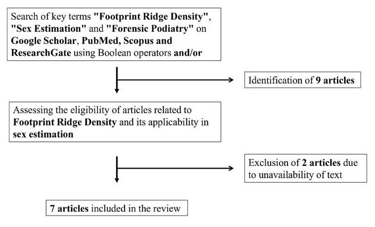
For the present mini review, the terms „Footprint Ridge Density”, „Sex Estimation” and „Forensic Podiatry” were searched on Google Scholar, PubMed, Scopus and ResearchGate as part of literature identification and screening using Boolean operators and/or. The inclusion criteria required articles that were written in English language and centered around FPRD, addressing its applicability in determining sex-based variability. Following the eligibility assessment, seven articles were reviewed that met the inclusion criteria between 2010 and August 2025, while two articles were excluded due to unavailability of the texts (Moorthy and Hairunnisa 2020, Moorthy et al. 2022). A flowchart depicting a schematic understanding of selecting the research articles is presented below (Fig. 1).
Footprint Ridge density (FPRD) analysis: Methodological overview
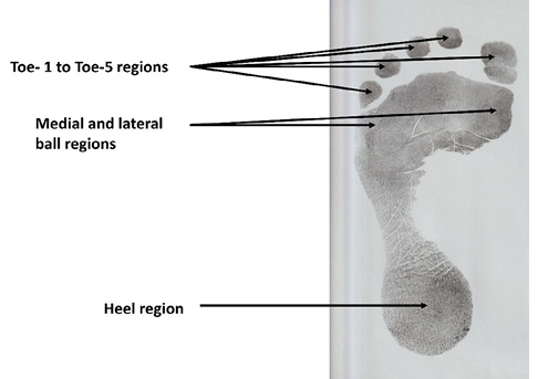
Ridge density quantification entails counting dermal ridges within predefined anatomical regions. It includes methodologies like measuring ridge counts between the core of the fingerprint patterns and the triradius (delta) (de Jongh et al., 2024) and between individual interdigital triradii in palmprints (Jerković et al., 2021). Similarly, Acree (1999) introduced a methodology to count ridges in fingerprints, which has become widely accepted in similar studies worldwide. He selected a predefined topological area of 5 × 5 mm outside the central core region of the radial side in bilateral fingers and diagonally counted the ridges to emphasize its variability due to differences in epidermal ridge thickness. This methodology was incorporated in the evaluation of FPRD by Kanchan et al. (2012) in a controlled environment. They examined four specific regions on the plantar surface of the foot to measure ridge density: at the medial side of the 1st toe, at the ball region of the 1st toe (also referred to as the medial ball), at the lateral ball region located beneath the triradius of the 5th toe, and at the central region of the heel. Moorthy and Hairunnisa (2018a) followed Acree’s (1999) methodology, but further expanded the number of measurable regions by incorporating the medial regions of each toe. Traditional studies analyzing ridge density patterns in footprints have focused on the depicted anatomical regions (see Fig. 2), with their statistical evaluations employing Receiver Operating Characteristic (ROC) and Area Under the Curve (AUC) analyses for the purpose of identifying robust indicators and their sex classification accuracy. More recently, Budka et al. (2021) revolutionized this traditional ridge count technique by replacing manual analysis with convolutional neural networks (CNNs) application to estimate sex from 2D footprint images. They formulated 10 mm² sampling squares from the entirety of a footprint, and developed learned models based on size, shape and texture of footprints that included incomplete or noisy ridge segments typical of ideal prints recovered from a crime scene.
Review of literature on sex estimation from FPRD
Kanchan et al. (2012) assessed the potential use of FPRD for sex estimation among 106 subjects from a medical college in Mangalore, Southern India, who were between the ages of 20 and 25. Findings inferred mean FPRD was 11.9 ridges/25 mm² or more for females and 10.4 ridges/25 mm² or less for males for medial ball region on the right side, and deemed most accurate region for sex determination, out of 212 footprints that were examined. Conversely, the heel area showed the least variation, with a FPRD value of 10.4 ridges/25 mm² or less was likely to be a male, and 11.0 ridges/25 mm² or more was considered as a female. The analyzed areas demonstrated statistically significant (p < 0.05) mean ridge density values, confirming the sex-based variation. Further, ROC curve analysis predicted medial ball region had the highest potential for sex discrimination, with an AUC value of 77.8% for the left foot and 86.5% for the right, followed by medial region of 1st toe, lateral ball and heel region. The researchers concluded that females have a higher ridge density than males, perhaps as a result of their finer ridges and narrower valleys in their dermatoglyphic patterns, showcasing the applicability of FPRD in estimating sex.
Sex differences from FPRD were examined by Krishan et al. (2015) among 160 college students of Shimla town in North India, aged 18 to 25 (39 males and 121 females). Based on the outcomes, females possessed a higher mean ridge density than males in each defined region of the footprints. The highest FPRD differences were observed in the medial ball region, where males showed a density value of 7.3 ridges/25 mm² or less, and females recorded a ridge density of 8.7 ridges/25 mm² or more. On the other hand, the heel region showed the least amount of variation between the sexes; for males, the discriminative accuracy was approximately 6.7 ridges/25 mm² or less, while for females, it was likely 7.5 ridges/25 mm² or more. On evaluating the sexing potential of ridge density through the ROC curve, right footprints demonstrated greater discriminatory power, and the medial ball region possessed the highest AUC of 85.3%, followed by 1st toe (81.7%), lateral ball (80.1%) and heel region (69.2%). The sexing potentials of the left and right footprints were 77.7% and 91.5%, respectively, according to ROC analyses of the total FPRD value.
Moorthy and Hairunnisa (2018a) conducted an investigation on the Bidayuh population of Malaysia (100 males and 100 females). They examined sex differences from FPRD in eight different areas among 400 footprints- medial regions of each toe, medial and lateral ball regions and heel region. Highest mean ridge density in males was found in 3rd and 4th toes (≤ 13.71 ridges/25 mm²), whereas females were identified by values of ≥ 14 ridges/25 mm². ROC curve analysis demonstrated strong sexing potential, with the right 1st toe yielding the highest AUC of 97.3%, followed by left 3rd toe (97.0%) and right medial ball (96.8%).
Moorthy and Hairunnisa (2018b) further evaluated toeless FPRD from the Iban ethnic group of Malaysia. The sample consisted of 200 individuals (100 males and 100 females) of age group 29–69 years. They analyzed ridge density in three regions: medial ball region, lateral ball region and heel region. Mean FPRD values showed medial ball region showed the highest discriminatory power for sex determination in the right side (male – 10.75 ridges/25 mm² or less; female – 12.18 ridges/25 mm² or more), followed by lateral ball region, while heel region demonstrated the lowest predictive strength. ROC analyses confirmed these findings, inflicting high sexing potential of FPRD in medial ball region (AUC – 94.8% for right side, 92.1% for left side), followed by lateral ball region (AUC – 95.7% for right side, 88.6% for left side) and heel region (AUC – 87.2% for right side, 92.6% for left side).
Moorthy and Hairunnisa (2019) examined sex differences from toeprint RD among 100 males and 100 females of the Melanau indigenous population in Malaysian Borneo Island. They found female ridge densities to be significantly higher in comparison to male ridge densities, indicating gender variation in all bilateral toes. For the left side, 3rd toe RD showed higher gender variation (t = 6.782), while for the right side, variations were recorded the highest in 4th toe (t = 8.689), followed by 3rd toe (t = 8.101).
Moorthy et al. (2021) analyzed sex variations from toeprint RD among the Kagay-Anon population of Cagayan de Oro city in Philippines; examining 4000 toe prints from 201 females and 199 males. ROC analyses determined the 3rd toe of the left side (AUC – 68.8%) and 4th toe of the right side have the highest sexing potential (AUC – 59.3%).
Budka et al. (2021) studied the sex estimation of the British population, aged 16 to 81, using texture density, size and shape of footprints. The researchers used CNNs to analyze two datasets: a pilot dataset of 196 footprints and a larger dataset of 2677 footprints. Their research highlighted the heel region, and the region between the 1st and 2nd toe as important contributors in discriminating sex. These regions were further validated by Grad-CAM heatmaps as being significant to the model’s decision-making process. The study demonstrated the potential of implementing algorithms trained with machine learning to identify sex by achieving an accurate prediction of nearly 83% on the larger dataset for texture analysis and ~ 90% upon including all the variables.
Discussion
Forensic assessments utilizing dermatoglyphic prints from the human body offer a cost-effective and non-invasive tool for human identification purposes. These prints exhibit inherent variations in ridge configurations, which form the basis for FPRD analysis (Kanchan et al., 2012). All the reviewed literature analyzed FPRD of populations with varied demographic backgrounds. While, Kanchan et al. (2012) studied young adults aged 20–25 years from Southern India, Krishan et al. (2015) considered students aged 18–25 years from Northern India. In contrast, Moorthy and Hairunnisa (2018a;b) researched on indigenous groups of Malaysia aged 29–69. Budka et al. (2021) further included British volunteers spanning a wide age range of 16–81 years, all demonstrating the validity of FPRD across diverse life stages while taking into consideration demographics that differ both genetically and environmentally.
The reviewed literature consistently highlighted significant sex-based differences in ridge density of footprints. For example, Kanchan et al. (2012) found females exhibited a mean FPRD of ≥ 11.9 ridges/25 mm² and males displayed a value of ≤ 10.4 ridges/25 mm² in the medial ball region of the footprint. Similarly, Moorthy and Hairunnisa (2018b) reported higher mean FPRD values in females (≥ 12.18 ridges/25 mm²) compared to males (≤ 10.75 ridges/25 mm²) also in the medial ball region. These outcomes are deemed similar to Acree’s (1999) preliminary research related to ridge density study of fingers, which established sex-specific ridge density thresholds of ≥ 12 ridges/25 mm² for females and ≤ 11 ridges/25 mm² for males. An analogous outcome was also observed in a study on the hypothenar region of palmprints, with females recording a mean ridge density of ≥ 13.5 ridges/cm², compared to ≤ 11.8 ridges/cm² for males (Kanchan et al., 2013). This parallel observation of higher ridge densities in females can be attributed to their finer ridge detailing and narrower valleys, as a characteristic of dermatoglyphic patterns across footprints, fingerprints and palmprints.
Moreover, variations in predictive capabilities across footprint regions highlights the need to generate unique models for sex estimation using FPRD. For example, medial ball region was identified as having higher discriminatory power for sex estimation in three reviewed studies (Kanchan et al., 2012; Krishan et al., 2015; Moorthy & Hairunnisa, 2018b), while Budka et al. (2021) highlighted heel regions as accurate most sex predictor. Heathfield et al. (2016) further supported the significance of heel region in their study to find out ethnic differences, comparing FPRD between two South African populations. The disparity indicates the population-specific nature of FPRD and the limitations of applying generalized prediction models universally, affirming one of the core principles of forensic anthropology. It also highlights the need of including every region in FPRD-based studies since it is very unlikely to predict which region of the footprint might be recovered from a crime scene. Table 1 summarizes the reviewed studies and lists accurate most regions of sex estimation from FPRD.
| Study reference | Demographic background | Sample size | Footprint regions | Most accurate sex predictor |
|---|---|---|---|---|
| Kanchan et al. (2012) | South Indian population | 56 males and 50 females | 1st toe, medial ball, lateral ball, heel | Medial ball |
| Krishan et al. (2015) | North Indian population | 39 males and 121 females | 1st toe, medial ball, lateral ball, heel | Medial ball |
| Moorthy and Hairunnisa (2018a) | Bidayuh ethnicity, Malaysian population | 100 males and 100 females | 1st–5th toe, medial ball, lateral ball, heel | Heel |
| Moorthy and Hairunnisa (2018b) | Iban ethnicity, Malaysian population | 100 males and 100 females | Medial ball, lateral ball, heel | Medial ball |
| Moorthy and Hairunnisa (2019) | Melanau ethnicity, Malaysian population | 100 males and 100 females | 1st–5th toe | 3rd toe, 4th toe |
| Budka et al. (2021) | British population | Pilot – 101 males and 132 females; Larger dataset – 1194 males and 1483 females | Toe regions, medial arch, lateral arch, ball of the foot, and heel | Heel, region between toes |
| Moorthy et al. (2021) | Kagay-Anons, Philippine population | 201 males and 199 females | 1st–5th toe | 3rd toe, 4th toe |
It is also evident from the studies reviewed that FPRD have shown promising percentages of accuracy in sex estimation, since Kanchan et al. (2012), Krishan et al. (2015) and Moorthy and Hairunnisa (2018b) reported medial ball region’s high discriminatory power with ROC analyses revealing AUC values of 86.5%, 85.3% and 94.8% respectively. These robust prediction rates align with similar investigations done on finger and palmar ridge densities. For instance, Qi et al. (2022) reported over 90% accuracy with supervised learning settings, while Das et al. (2024) achieved an accuracy exceeding 80% using traditional methods, both from fingerprints. A success rate over 80% was also reported in research on palmprint ridges analysis (Gutiérrez-Redomero & Alonso-Rodríguez, 2013). Comparisons thus infer FPRD can be seen as a reliable and consistent biometric parameter to establish sex differences.
Technological advancements like the utilization of Artificial Intelligence (AI) algorithms underscore the potential to extract meaningful footprint patterns and establish sex-based variability from ridge analyses (Budka et al., 2021). This progress aligns with the broader biometric application of AI in ridge density analyses of other body prints, as noted by the use of autoencoder networks on fingerprints (Qi et al., 2022).
Limitations and future applications
Numerous studies exist that have examined morphometric characters of footprints to estimate sex. However, research related to quantification of plantar ridges is still at an initial level. A key limitation in the majority of existing studies till date is the uptake of smaller sample size and lacking consideration of environmental variations in previous studies, which may restrict the generalizability of the findings. Thus, the present mini review voices for potential use and standardization of FPRD as a parameter for sex identification. It highlights the necessity to develop population and region-specific sex estimation algorithms using larger samples in different forensic casework scenarios, as existing literature indicates substantial threshold values of FPRD (Kanchan et al., 2012; Krishan et al., 2015).
Traditional dermatoglyphic techniques for analyzing FPRD provide direct and interpretable observations while being comparatively inexpensive to capture fine-scale ridge features without computational requirements. However, these methods are susceptible to inter-observer variability, labor-intensiveness, and also challenging to scale for larger repositories. Thus, the present study suggests for complementing the integration of AI in sex estimation studies on FPRD, rather than replacing traditional methodologies, as generating supervised models using Machine Learning (ML) and Deep Learning (DL) algorithms can reduce observer bias, improve reproducibility and enhance predictive capability for sex estimation to analyze complex datasets (Budka et al., 2021; Kanchan et al., 2012).
Future studies can investigate the relationship between FPRD and other biological profiling factors, such as body weight and stature, and broaden its application beyond sex estimation. Examining these relationships may yield profound understanding of the trends in ridge count variability and how it relates to physical characteristics of the human body. Further explorations on tracing ancestry through FPRD is also recommended. Although previous studies have demonstrated the value of ethnicity profiling from FPRD (Heathfield et al., 2016), carrying out comparable studies in other populations could contribute to an improved understanding of this aspect.
Since research within the domain of fingerprinting has established correlations between ridge density and epidemiological attributes like diabetes, hypertension and genetic anomalies (Sharma et al., 2021), implementing these outcomes can open opportunities for investigations into the variables affecting sexual variations in FPRD. Expanding this research area thus can help establish links between dermatoglyphic traits of footprints and underlying genetic or hormonal factors. Additionally, studying the influence of occupational factors might offer another direction for future investigations, as in many circumstances, people of rural and environmentally secluded communities travel barefooted for socio-economic and occupational reasons, it can impact the pattern of plantar ridges over time (Krishan, 2008).
Conclusion
The present mini review concludes that FPRD is a relatively novel parameter that has the potential of contributing to sex estimation studies. Its application not only offers an upgradation on the existing value of footprints in forensic podiatry, but also promises valuable contributions to broader fields of human genetics and anthropological sciences.
References
Acree, M. A. (1999). Is there a gender difference in fingerprint ridge density? Forensic Science International, 102(1), 35–44. https://doi.org/10.1016/S0379-0738(99) 00037-7
Ali, F. I., & Ahmed, A. A. (2020). Sexual and topological variability in palmprint ridge density in a sample of Sudanese population. Forensic Science International: Reports, 2, 100151. https://doi.org/10.1016/j.fsir.2020.100151
Babler, W. J. (1991). Embryologic development of epidermal ridges and their configurations. Birth Defects Original Article Series, 27(2), 95–112.
Budka, M., Bennett, M. R., Reynolds, S. C., Barefoot, S., Reel, S., Reidy, S., & Walker, J. (2021). Sexing white 2D footprints using convolutional neural networks. Plos one, 16(8), e0255630. https://doi.org/10.1371/journal.pone.0255630
Campo, E. (2018). Exploring various techniques to process and identify latent friction ridge details from fingers on semi-porous surfaces. Honors thesis: 567. University of Southern Mississippi. Available at: https://aquila.usm.edu/honors_theses/567/ [Accessed 2 November 2025].
Champod, C., & Evett, I. W. (2001). A probabilistic approach to fingerprint evidence. Journal of Forensic Identification, 51(2), 101.
Cummins, H., & Midlo, C. (1926). Palmar and plantar epidermal ridge configurations (dermatoglyphics) in European-Americans. American Journal of Physical Anthropology, 9(4), 471–502. https://doi.org/10.1002/ajpa.1330090422
Das, D., Seal, S., Pal, S., Chitara, N., Meena, R., Guleria, A., Rana, A., Verma, R. & Krishan, K. (2024). Sexual dimorphism and topological variability in fingerprint ridge density in a north-west Indian population. The Science of Nature, 111(3), 23. https://doi.org/10.1007/s00114-024-01911-x
de Jongh, A., Lubach, A. R., Lie Kwie, S. L., Loadsman-Wammes, F. D., & Alberink, I. (2024). Measuring the rarity of core-delta distances in fingerprint patterns in the Dutch population. Journal of Forensic Sciences, 69(1), 94–116. https://doi.org/10.1111/1556-4029.15381
Dhall, J. K., & Kapoor, A. K. (2016). Fingerprint ridge density as a potential forensic anthropological tool for sex identification. Journal of Forensic Sciences, 61(2), 424–429. https://doi.org/10.1111/1556-4029.12959
Dhaneshwar, R., Kaur, M., & Kaur, M. (2021). An investigation of latent fingerprinting techniques. Egyptian Journal of Forensic Sciences, 11(1), 33. https://doi.org/10.1186/s41935-021-00252-4
Gutiérrez-Redomero, E., Alonso, C., Romero, E., & Galera, V. (2008). Variability of fingerprint ridge density in a sample of Spanish Caucasians and its application to sex determination. Forensic Science International, 180(1), 17–22. https://doi.org/10.1016/j.forsciint.2008.06.014
Gutiérrez-Redomero, E., & Alonso-Rodríguez, C. (2013). Sexual and topological differences in palmprint and ridge density in the Caucasian Spanish population. Forensic Science International, 229(1–3), 159–e1. https://doi.org/10.1016/j.forsciint.2013.03.014
Gutiérrez-Redomero, E., Quirós, J. A., Rivaldería, N., & Alonso, M. C. (2013). Topological variability of fingerprint ridge density in a sub-saharan population sample for application in personal identification. Journal of Forensic Sciences, 58(3), 592–600. https://doi.org/10.1111/1556-4029.12092
Heathfield, L. J., Prins, A. M., & Schall, R. (2016). Comparison of footprint ridge density between two South African ethnic groups. J Forensic Investigation, 4(1), 4. https://doi.org/10.13188/2330-0396.1000031
Jerković, I., Ljubić, T., Bardić, L., Kolić, A., & Anđelinović, Š. (2023). Application of palmar digital intertriradial distances for sex classification from palmprints: a preliminary study. Australian Journal of Forensic Sciences, 55(1), 34–45. https://doi.org/10.1080/00450618.2021.1882573
Kanchan, T., Krishan, K., Aparna, K. R., & Shyamsunder, S. (2012). Footprint ridge density: a new attribute for sexual dimorphism. Homo, 63(6), 468–480. https://doi.org/10.1016/j.jchb.2012.09.004
Kanchan, T., Krishan, K., Aparna, K. R., & Shyamsundar, S. (2013). Is there a sex difference in palm print ridge density? Medicine, Science and the Law, 53(1), 33–39. https://doi.org/10.1258/msl.2012.011092
Kapoor, N., & Badiye, A. (2015). Sex differences in the thumbprint ridge density in a central Indian population. Egyptian Journal of Forensic Sciences, 5(1), 23–29. https://doi.org/10.1016/j.ejfs.2014.05.001>
Krishan, K. (2008). Estimation of stature from footprint and foot outline dimensions in Gujjars of North India. Forensic Science International, 175(2–3), 93–101. https://doi.org/10.1016/j.forsciint.2007.05.014
Krishan, K., Kanchan, T., Sharma, R., & Pathania, A. (2014). Variability of palmprint ridge density in a North Indian population and its use in inference of sex in forensic examinations. Homo, 65(6), 476–488. https://doi.org/10.1016/j.jchb.2014.08.003
Krishan, K., Kanchan, T., Pathania, A., Sharma, R., & DiMaggio, J. A. (2015). Variability of footprint ridge density and its use in estimation of sex in forensic examinations. Medicine, Science and the Law, 55(4), 284–290. https://doi.org/10.1177/0025802414557880
Liu, L. (2025). Types and distribution of the friction ridge patterns on the ball area of the bare footprint. Forensic Sciences Research, 10(3), owaf015. https://doi.org/10.1093/fsr/owaf015
Malik, H. M. A., & Bashir, K. (2023). The detection and identification of footprint impressions at the scene of crime – A mini review. Forensic Insights and Health Sciences Bulletin, 1(2), 44–49. https://doi.org/10.56770/fi2023113
Mohamed, K. M., Gaballah, E. M. F., Zaghloul, H. S., & Ismail, M. M. (2020). Palm ridge density as anew identifier for age and sex in forensic medicine. QJM: An International Journal of Medicine, 113(Supplement_1), hcaa049–003. https://doi.org/10.1093/qjmed/hcaa049.003
Moorthy, T. N., & Hairunnisa, M. A. K. (2018a). Sex determination from footprint ridge density in bidayuh population in Malaysian Borneo. International Journal of Medical Toxicology & Legal Medicine, 21(3–4), 158–161. https://doi.org/10.5958/0974-4614.2018.00057.8
Moorthy, T. N., & Hairunnisa, M. A. K. (2018b). Gender variation from footprint toes among ibans, An indiginenious ethnic group in Malaysian Borneo. International Journal of Medical Toxicology & Legal Medicine, 21(3–4), 137–140. https://doi.org/10.31031/FSAR.2018.03.000577
Moorthy, T. N., & Hairunnisa, M. (2019). Gender variation from toe print ridge density among Melanau ethnic in Malaysia Borneo Island. International Journal of Medical Toxicology & Legal Medicine, 22(1–2), 8–12. https://doi.org/10.5958/0974-4614.2019.00003.2
Moorthy, N. T., & Hairunnisa, M. A. K. (2020). Gender variation from footprint ridge density among Lun Bawangs of Malaysian Borneo for crime scene application. International Journal of Medical Toxicology & Legal Medicine, 23(3–4), 61–66. https://doi.org/10.5958/0974-4614.2020. 00047.9
Moorthy, T. N., Devina, K. D., Nikkimor, I. L. D. & Philip, A. I. P. (2021). Gender determination from toe prints among Kagay-Anons of Philippines for forensic application. Indian Journal of Forensic Medical Toxicology 15(2), 1125–1130.https://doi.org/10.37506/ijfmt.v15i2.14470
Moorthy, T. N., Syarani, N., & Pritam, H. M. H. (2022). Sexual dimorphism from toe prints among Malaysian Malays for person identification. Journal of Krishna Institute of Medical Sciences (JKIMSU), 11(1).
Naffah, J. (1977). Dermatoglyphic analysis: anthropological and medical aspects. Bulletin of the New York Academy of Medicine, 53(8), 681.
Nayak, V. C., Rastogi, P., Kanchan, T., Yoganarasimha, K., Kumar, G. P., & Menezes, R. G. (2010). Sex differences from fingerprint ridge density in Chinese and Malaysian population. Forensic Science International, 197(1–3), 67–69. https://doi.org/10.1016/j.forsciint.2009.12.055
Patnaik, B. B., Penmetsa, G. S., Vinnakota, K., Ramaraju, A. V., & Alla, R. K. (2024). Identification of dermal crease patterns as a link between genetics and periodontitis: reliability and credibility. Journal of Forensic Science and Medicine, 10(2), 106–110. https://doi.org/10.4103/jfsm.jfsm_70_23
Polcerová, L., Chovancová, M., Králík, M., Beňuš, R., Klíma, O., Meinerová, T., Čuta, M., & Petrová, M. E. (2022). Radioulnar contrasts in fingerprint ridge counts: Searching for dermatoglyphic markers of early sex development. American Journal of Human Biology, 34(5), e23695. https://doi.org/10.1002/ajhb.23695
Polcerová, L., Jantz, R. L., Králík, M., Chovancová, M., & Čuta, M. (2023). Sex differences in radioulnar contrasts of the finger ridge counts across 21 human population samples. Annals of human biology, 50(1), 370–389.https://doi.org/10.1080/03014460.2023.2247970
Qi, Y., Qiu, M., Jiang, H., & Wang, F. (2022). Extracting fingerprint features using autoencoder networks for gender classification. Applied Sciences, 12(19), 10152. https://doi.org/10.3390/app121910152
Reichardt, G. J., Carr, J. C., & Stone, E. G. (1978). A conventional method for lifting latent fingerprints from human skin surfaces. Journal of Forensic Sciences, 23(1), 135–141. https://doi.org/10.1520/JFS10662J
Schaumann, B., & Alter, M. (1976). Embryogenesis and genetics of epidermal ridges. Dermatoglyphics in medical disorders, 1–11. Berlin, Heidelberg: Springer Berlin Heidelberg. https://doi.org/10.1007/978-3-642-51620-7_1
Sharma, S., Shrestha, R., Krishan, K., & Kanchan, T. (2021). Sex estimation from fingerprint ridge density. A review of literature. Acta Bio Medica: Atenei Parmensis, 92(5), e2021366. https://doi.org/10.23750/abm.v92i5.11471
Soanboon, P., Nanakorn, S., & Kutanan, W. (2016). Determination of sex difference from fingerprint ridge density in northeastern Thai teenagers. Egyptian Journal of Forensic Sciences, 6(2), 185–193. https://doi.org/10.1016/j.ejfs.2015.08.001
Final information
Contributions from individual authors
SS: Conceptualization, data collection, drafting of manuscript, data analysis, data interpretation, final version approval; MS: data collection, drafting of manuscript, data analysis, data interpretation, final version approval; MC: drafting of manuscript; review & editing, final version approval; PŠ: drafting of manuscript, review & editing, final version approval, manuscript supervision; RB: drafting of manuscript, review & editing, final version approval, manuscript supervision
Ethics statement
Not applicable as this is a literature review.
Data availability statement
No raw data were generated in this study. The reviewed articles are referenced in text.
Financial disclosure
None to declare.
Conflict of Interest
None to declare.
Corresponding Author
Mr. Saumya Seal, M.Sc., Ph.D. Student, Department of Anthropology, Faculty of Natural Sciences, Comenius University, Ilkovičova 6, Mlynská dolina, 842 15 Bratislava 4, Slovak Republic, e-mail: seal1@uniba.sk