Frequency of Basic Types of Dorsal Hand Vein Patterns in the Slovak Population
 https://orcid.org/0000-0001-7497-4359
Department of Anthropology, Faculty of Natural Sciences, Comenius University in Bratislava, Bratislava, Slovakia
https://orcid.org/0009-0006-0125-9673
Department of Anthropology, Faculty of Natural Sciences, Comenius University in Bratislava, Bratislava, Slovakia
Department of Anthropology, Faculty of Natural Sciences, Comenius University in Bratislava, Bratislava, Slovakia
https://orcid.org/0000-0003-1114-124X
Department of Anthropology, Faculty of Natural Sciences, Comenius University in Bratislava, Bratislava, Slovakia
Department of Anthropology, Faculty of Natural Sciences, Comenius University in Bratislava, Bratislava, Slovakia
https://orcid.org/0000-0001-5917-1689
Department of Anthropology, Faculty of Natural Sciences, Comenius University in Bratislava, Bratislava, Slovakia
Abstract
Introduction
Dorsal hand vein pattern represents a unique morphological feature of the human body which may serve as a biometric tool for forensic identification.
Study Aim
The primary aim of this study was to determine the frequency and distribution of dorsal hand vein patterns in a Slovak adult population, with respect to sex and laterality of the hand.
Material and Methods
This study provides a morphological analysis of dorsal hand vein patterns in a sample of 70 healthy adults from the Slovak population. Vein configurations were classified using the 1951 system developed by Suchý, distinguishing four main types: branched, double-branched, simple, and composite.
Results
The most frequent patterns were branched and double-branched, while the composite form was rare. No statistically significant differences were found between sexes or between hands, suggesting a high degree of bilateral and intersexual symmetry. A rare morphological subtype, labelled 2N4, appeared exclusively in females on the left hand, potentially reflecting sex-linked vascular variation.
Conclusion
The results support the hypothesis that dorsal venous architecture is largely determined by early developmental and genetic factors. Given the pattern stability and inter-individual variability, dorsal hand veins remain a promising biometric marker. Despite limitations related to imprinting technique and assessment subjectivity, the study offers a valuable anatomical reference for future biometric, forensic, or anthropological research.
Keywords: venous morphology, biometric identification, sex differences, vascular symmetry
Introduction
The dorsal hand vein pattern represents a unique morphological feature of the human body, which has become a subject of interest in recent decades for researchers in the fields of biometric systems and clinical practice. This pattern is formed by a network of metacarpal and dorsal veins located beneath the skin, creating an individual-specific pattern (Elmegarhi et al., 2018). Due to its high level of uniqueness and stability over a lifetime, dorsal hand vein patterns have emerged as a valuable tool in biometric identification, forensic analysis, and medical diagnostics (Hsu et al., 2011). The dorsal hand vein pattern shows significant individual variability, resulting from a combination of genetic predispositions and external factors. The characteristic pattern is formed by the main metacarpal veins, often complemented by finer branches known as minutiae. These small veins may connect the main branches, encircle them, or form independent structures, contributing to the uniqueness of individual’s vein pattern (Elmegarhi et al., 2018; Suchý, 1951a).
Research suggests that the basic type of vein pattern is genetically determined, with individuals possessing a thicker subcutaneous fat layer more frequently exhibiting a less complex and simpler vein pattern (Hung et al., 2022; Suchý, 1951b). However, the genetic basis of variability in dorsal hand vein patterns remains underexplored. The first study was conducted by Suchý (1951b) on a sample of 66 people, including 13 complete families. The results showed similarities in vein patterns not only among siblings but also across generations.
The visibility of the vein pattern is influenced by both external and individual factors. The prominence of veins increases with age due to the natural reduction of subcutaneous fat on the dorsal hand, making both veins and tendons more visible (Hung et al., 2022). Additionally, medical conditions such as long-term intravenous infusions can lead to thrombosis and subsequent vein occlusion, further altering the pattern over time (Fiala et al., 2015). Physical labor and sports activities also increase vein visibility, especially in simple vein patterns, while environmental temperature causes veins to appear bluish in cold and more prominent in warm conditions (Suchý, 1951b; Hung et al., 2022). Sexual dimorphism has been observed, with males generally exhibiting more pronounced veins, particularly in physically active individuals, but these differences diminish with age. The visibility of vein relief is also influenced by factors that affect vasoconstriction or vasodilatation. Vasoconstriction is the process of contracting smooth muscle tissue in the blood vessel walls, narrowing the vessels and increasing the blood pressure, making veins less visible. Factors with vasoconstrictive effects include hormones (norepinephrine, vasopressin), medications and drugs (pseudoephedrine, corticosteroids, nicotine) and dietary substances (caffeine, salt, licorice). On the contrary, vasodilatation causes relaxation of smooth muscles in the vein walls, widening their diameter and increasing blood flow, making veins more prominent and visible. Such factors include prostaglandins, adenosine, histamine, and dietary substances such as garlic, dark chocolate and regular physical activity (Charkoudian, 2010; Hung et al., 2022; Suchý, 1951b; Zrzavý, 1957).
Asymmetry of the vein pattern is another important feature, initially linked to motor dominance, although later studies did not confirm this view (Černáček, 1994; Kaczeńska & Dilling-Ostrowska, 1961; Minor, 1931). Freerksen (1938) found asymmetry between the right and left hands of monozygotic twins, and Suchý (1961) defined four degrees of relative symmetry, with absolute bilateral symmetry occurring in only 0.2% of cases.
In recent years, research on vein patterns has expanded to their use in biometric applications, identification systems, and forensic sciences (Hartung et al. 2020; Wilson 2010). Hartung et al. (2020) demonstrated that dorsal hand vein patterns are sufficiently individual for reliable person identification, with the probability of a random match between two people being less than 1:1000. Their study analyzed vein patterns of 30 participants using standardized grid analysis, quantifying the number of vein crossings and branching in different hand regions. The results indicated a high stability of vein patterns, supporting their use in identification systems. Furthermore, the study highlighted the existence of a “region with reduced vein density” between the proximal halves of the second and third metacarpal bones, where vein density is lower, which may affect identification accuracy in certain areas. Hartung et al. (2020) also noted that factors such as age, sex, and health condition can influence vein visibility and pattern, with veins being more prominent in older individuals and those with low subcutaneous fat.
The vein pattern is immutable, non-transferable, and characterized by high accuracy (Ahmed et al., 2013). In contrast to other biometric features, such as papillary terrain or the iris, which may be sensitive to age-related structural changes, vein pattern identification offers higher security since veins lie deeper under the skin and are less prone to potential forgery (Harchana et al., 2021). Vein pattern imaging systems use infrared diodes and scanners with weather shields, allowing for accurate visualization of veins as the warmest objects in the image. The procedure is non-invasive and easy to perform under standardized conditions with an error rate of just 0.01% (Rak et al., 2018; Wilson, 2010). Unlike face or iris scanning, which may be influenced by external factors such as lighting or wearing glasses, vein patterns provide more stable and reliable results in various operational conditions. Vein pattern technology thus meets all the main characteristics of a good biometric trait: universality, uniqueness, permanence, ease of collection, high accuracy, and wide acceptance worldwide (Harchana et al., 2021).
The primary aim of this study was to determine the frequency and distribution of dorsal hand vein patterns in a Slovak adult population, with respect to sex and laterality of the hand. The research aims to deepen the understanding of anatomical variability and assess the potential forensic and biometric applications of vein pattern analysis. By enhancing knowledge in this field, the study seeks to contribute to the development of more accurate and secure identification systems for forensic and security purposes.
Material and Methods
This study examined a total of 70 participants from Slovakia, consisting of 33 females and 37 males, with an age range of 18 to 83 years and an average age of 39.49 years. Participation was voluntary and written informed consent was obtained. The consent included details about the study, such as its aims, methodology and other participation information. Withdrawal from the study was possible at any time. For each participant, the following information was recorded: identification number, sex, age, and any familial relationship with other participants. Imprints of the dorsal hand veins were obtained from both the right and left hands of each participant. To document the dorsal hand vein patterns, a method described by Suchý (1951b) was applied. This method involves taking imprints of the superficial veins on the dorsal surface of the hand and is practical, fast, and applicable in various settings. However, it may be influenced by subjective error, especially in individuals with a thicker layer of subcutaneous fat.
The equipment used included an Esmarch bandage, oily cream, a fine brush, stamp ink diluted with water at a ratio of 1:50, filter paper (135 mm x 85 mm), a pencil, as well as cotton and cleaning agents such as benzine and warm water. The procedure to enhance vein visibility included several techniques: compression of the veins on the palmar side of the forearm using the middle three fingers with the thumb resting on the ulna approximately 5 cm from the olecranon ulnae compression at the level of the processus styloideus radii and processus styloideus ulnae with simultaneous dynamic finger exercises (alternating finger spreading and fist clenching); application of an Esmarch bandage above the elbow combined with wrist movements; application of an Esmarch bandage below the elbow with finger exercises; immersion in warm water; and rapid immersion in cold water (Suchý, 1951b).
Subsequently, the dorsal surface of the hand was coated with oily cream in the metacarpal and carpal areas, and ink was applied along the visible veins using a brush in the direction from the fingers. The vein paths were marked while the participant’s hand was clenched into a fist to minimize the influence of factors such as physical activity, sports, or age. The vein patterns were then transferred by pressing filter paper onto the inked dorsal surface of the clenched hand. The paper was placed perpendicular to the longitudinal axis of the hand, with the label positioned on the radial side. Finally, the interphalangeal joint spaces were numbered with a pencil directly on the hand, starting from the thumb (marked as one) to the little finger (marked as five).
Evaluation of Vein Patterns
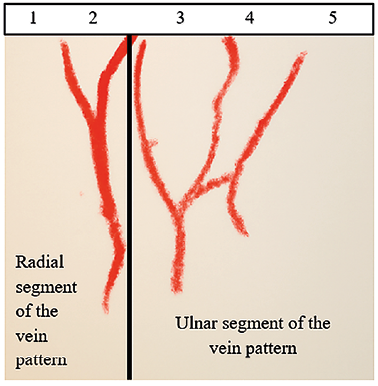
The dorsal hand vein system was assessed according to the methodology of Suchý (1951b). The evaluation included classification based on the ratio of radioulnar branch distribution and the branching type of the ulnar segment. The venous formula was determined according to Suchý’s numerical system (1961). The radioulnar segments of the dorsal venous network are typically composed of an ulnar segment and a radial segment (Figure 1). The ulnar segment usually originates from the second, third, or fourth interphalangeal spaces and contributes significantly to the venous pattern. The radial segment predominantly drains the thumb and radial side of the index finger and often includes a prominent branch arising from the second interphalangeal space. Each segment has a principal vein trunk. In the ulnar segment, this is typically a branch from one of the mentioned interphalangeal spaces, while in the radial segment, the main trunk most commonly originates from the second interphalangeal space or the radial side of the index finger if no connection to this space is present. In rare cases, the pattern also includes the vena basilica when it connects to the ulnar trunk of the vena cephalica before crossing the limiting line defined by the so-called limiting line of the dorsal venous pattern, which runs between the processus styloideus radii and processus styloideus ulnae (Suchý 1951b).
Vein patterns were classified visually by one rater according to the criteria described by Suchý (1951). The classification was based on direct visual comparison of the imprints with defined morphological types. The assessment was not blinded, as the rater had access to demographic and hand-side information during evaluation. Demographic data are essential for interpreting responses accurately, as the data access to demographics allows to assess whether responses align with expected norms. Interobserver reliability was not tested, as all patterns were evaluated by a single examiner, trained to recognize and minimize personal biases. These aspects may represent methodological limitations and should be considered when interpreting the results.
The decision to use the classification system described by Suchý (1951) was based on its historical relevance and frequent application in Central European anthropological studies. Despite the existence of modern, image-based classification systems, the Suchý method (1951) remains suitable for visual evaluation from ink-based imprints, which was the chosen technique in this study.
Additionally, the use of ink transfer may have introduced minor artifacts such as smearing, uneven pressure, or incomplete contact with the skin, potentially affecting the accuracy of recorded vein patterns. These limitations were minimized through standardized procedures but cannot be entirely excluded.
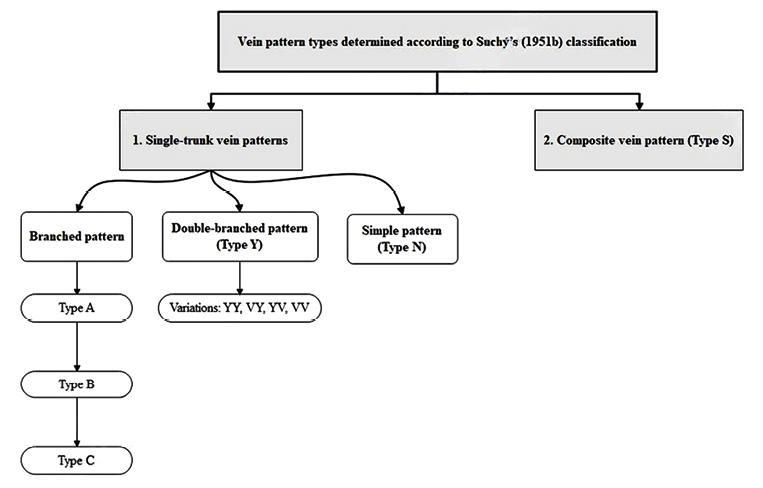
Vein pattern types were determined based on Suchý’s (1951b) classification system (Figure 2), which divides dorsal hand vein patterns into two primary categories: single-trunk vein patterns and composite vein patterns.
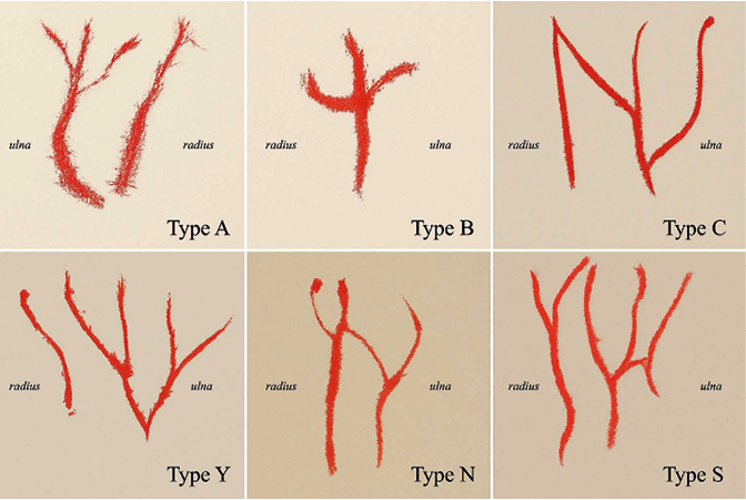
Single-trunk vein patterns include three subtypes (Figure 3):
- Branched pattern – three veins converge: the main trunk arises from the third space and receives branches from the second and fourth spaces. Variants include: Type A–the branch from the fourth space drains into the main trunk more distally than the branch from the second space; Type B–branches from the second and fourth spaces drain at approximately the same level; and Type C– the branch from the fourth space drains proximally relative to the branch from the second space.
- Double-branched pattern (Type Y) – the main trunk splits into two venous arms, each forming a Y-shaped bifurcation, resembling the letters “YY.” Variations such as VY, YV, and VV may occur.
- Simple pattern (Type N) – composed of two branches originating from either the second and third, third and fourth, or second and fourth spaces. The overall pattern resembles the letter “N” or “H”.
The second main category is the composite vein pattern (Type S), where branches originate from the radial trunk of the vena cephalica, often extending into the area of the second interphalangeal space.
Statistical Analysis
The observed vein pattern types were evaluated as qualitative traits, and their frequencies in the study population were expressed as percentages (p). For each percentage value, the standard error (sp) was calculated using the formula:
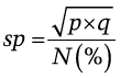
Where p = observed percentage, q = 100 − p, N = total number of observations.
To assess the significance of differences between frequencies, Fisher’s exact test and the chi-square test were used. Statistical calculations were performed using online calculators available at www.graphpad.com and www.quantpsy.org. The results were compared with critical values at a 5% significance level. A p-value of less than 0.05 was considered statistically significant.
Results
A total of 140 dorsal hand vein imprints (from 70 individuals; 66 female and 74 male hands) were analyzed, with imprints taken from both the left and right hands. Radioulnar segments of the dorsal venous network were generally well developed. In 98.57% of individuals, both the radial and ulnar parts of the vein pattern were simultaneously present on the dorsal surface of the hands. Only one participant (1.43%) exhibited a vein pattern consisting solely of the ulnar component.
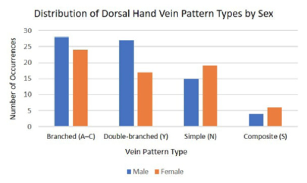
Across both hands and sexes, the most frequently observed pattern was the branched configuration (A–C), found in 37.14% of all cases. This was followed by the double-branched (Type Y) pattern with a frequency of 31.43%, and the simple (Type N) pattern, occurring in 24.29% of cases. The composite (Type S) vein pattern was rare, comprising only 7.14% of the total sample (Table 1).
Vein pattern frequencies varied slightly between sexes. For the right hand, the simple pattern (N) occurred in 33.33% of females and 21.62% of males (p = 0.30), while the branched pattern (A–C) appeared in 39.39% of females and 29.73% of males (p = 0.86). On the left hand, the double-branched pattern (Y) was present in 30.30% of females and 32.43% of males (p = 0.20). Across all types, inter-sex differences were not statistically significant (Table 1, Figure 4).
No statistically significant bilateral differences were observed between the right and left hands for any vein pattern type in either sex. In males, the most noticeable asymmetry appeared in the branched pattern, which was more frequent on the left hand, while in females, the double-branched pattern showed a slightly higher prevalence on the left. For the composite pattern, minor side differences were noted in both sexes. However, none of these differences reached statistical significance, with bilateral p-values ranging from 0.23 to >0.99 (Table 1), indicating an overall symmetry in vein pattern distribution.
Vein formulae were established for each hand using Suchý’s classification system (1951b, 1961), taking into account the origins and connections of individual vein branches. Most individuals showed symmetrical or near-symmetrical formulae between their right and left hands, although complete symmetry (identical formulae) was rare.
| Sex | Male (M) | *p | Female (F) | **p | Total | p-value (M vs. F) | ||
|---|---|---|---|---|---|---|---|---|
| Hand | right | left | right | left | ||||
| Branched (A–C) | 11 | 17 | 0.23 | 13 | 11 | 0.80 | 52 | 0.863 |
| % | 29.73 | 45.95 | 39.39 | 33.33 | 37.14 | |||
| Double-branched (Y) | 15 | 12 | 0.63 | 7 | 10 | 0.57 | 44 | 0.204 |
| % | 40.54 | 32.43 | 21.21 | 30.30 | 31.43 | |||
| Simple (N) | 8 | 7 | >0.99 | 11 | 8 | 0.59 | 34 | 0.324 |
| % | 21.62 | 18.92 | 33.33 | 24.24 | 24.29 | |||
| Composite (S) | 3 | 1 | 0.61 | 2 | 4 | 0.67 | 10 | 0.516 |
| % | 8.11 | 2.70 | 6.06 | 12.12 | 7.14 | |||
| Total | 37 | 37 | 33 | 33 | 140 | |||
*p-value: bilateral differences of basic dorsal hand vein pattern types in males
**p-value: bilateral differences of basic dorsal hand vein pattern types in females
p-value (M vs. F): differences in vein pattern types between males and females (combined data from both hands)
Discussion
This study provides a detailed overview of the frequency and distribution of basic dorsal hand vein pattern types in a sample of the Slovak adult population. The most common patterns observed were the branched type (A–C) and the double-branched type (Y), followed by the simple type (N). The composite pattern (S) was found to be the least prevalent. These findings are consistent with the notion that the venous architecture of the dorsal hand typically follows a branching morphotype, allowing for efficient drainage through multiple interconnected pathways.
Although slight differences in the frequency of certain patterns were observed between males and females, none of these differences reached statistical significance. For example, the simple pattern (N) appeared more frequently in females than in males, particularly on the right hand (33.33% vs. 21.62%; p = 0.30), while the branched pattern was more common in males. However, Fisher’s exact test revealed that these sex-related differences were not statistically significant (p = 0.204–0.863). These findings suggest that sex may not play a major role in determining the morphotype of dorsal hand vein patterns, at least within this sample.
Interestingly, a specific morphological variant of the simple type, labelled as 2N4 (indicating origin from the second and fourth interphalangeal depressions), was observed exclusively in females and only on the left hand. While the frequency of this pattern was low, its localized appearance may suggest an underlying anatomical or developmental predisposition, potentially influenced by sex-linked vascular variation. These finding merits further investigation in larger or age-stratified cohorts.
The high occurrence of the 2N4 type in the analyzed Slovak population may be related to specific developmental, genetic, or population-based factors. Several studies suggest that dorsal hand vein patterns are stable, unique, and likely influenced by early vascular development and inherited traits (Alashik & Yildirim, 2021; Hsu et al., 2011).
Some infrared imaging studies indicate that dorsal vein morphology exhibits inter-individual variability, which may also reflect underlying demographic or population-level differences (Hartung et al., 2020; Wang et al., 2021).
From a biometric perspective, the high frequency and localized presence of the 2N4 type may enhance recognition accuracy if included in algorithmic templates. Recent studies using deep learning and vein segmentation techniques, for example graph-based pattern matching or U-Net variants, have confirmed that regular, well-defined vein configurations significantly improve biometric performance (Lefkovits et al., 2022; Lajevardi et al., 2014).
Regarding bilateral distribution, no significant differences were found between the right and left hands within individuals. Frequencies of vein pattern types on both sides were similar across the population. The branched and double-branched types together accounted for more than 68% of patterns on each hand, with minor variations. Statistical analysis did not reveal significant bilateral asymmetry (e.g., p = 0.23 for branched type in males; p = 0.80 in females), with bilateral p-values ranging from 0.23 to >0.99 in males and from 0.57 to 0.80 in females (Table 1).
Vein pattern formulae were established for each hand using Suchý’s classification system (1951b; 1961), taking into account the origins and connections of individual vein branches. Most individuals showed symmetrical or near-symmetrical formulae between their right and left hands, although complete symmetry (identical formulae) was rare. This variability may reflect functional adaptation or minor individual anatomical variation.
The high prevalence of both radial and ulnar components in 98.57% of participants further confirms the typical bipartite organization of dorsal hand venous networks. The single case exhibiting only the ulnar component could represent a natural anatomical variant or an underdeveloped radial segment, but given its rarity, no general conclusion can be drawn from this observation.
Although this study focused on a morphological classification using anatomical imprint analysis, several recent works have demonstrated the utility of imaging-based and biometric approaches. For instance, Hsu et al. (2014) applied Gaussian directional filters to infrared images to enhance vein patterns for biometric recognition, achieving robust performance (Hsu et al., 2014). Similarly, Huang et al. (2015) combined vascular and subcutaneous tissue features for hand dorsum identification using multi-source key points and vein textures (Huang et al., 2015). While these approaches differ methodologically, they reinforce the anatomical assumption of individual uniqueness and bilateral symmetry in dorsal venous architecture. This supports the relevance of our findings, even when interpreted through modern biometric frameworks.
In recent years, research in the field of dorsal hand vein biometrics has focused on the use of advanced technologies such as infrared (IR) imaging and deep learning. These methods allow for more precise capture and analysis of vein patterns, thereby increasing the reliability of identification systems.
For example, the study by Hsu et al. (2011) presents an approach to personal authentication using dorsal vein patterns through infrared imaging, achieving high identification accuracy. Further research by Nayebi and Turgut (2021) demonstrates the effectiveness of deep neural networks in classifying vein patterns, reaching an accuracy of up to 99.64%. These studies confirm the potential of modern technologies in the field of vein pattern biometrics.
There are several limitations that should be acknowledged. First, the sample size, while sufficient for exploratory analysis, may not allow the detection of small but meaningful differences, especially in less common pattern types such as the composite form. It should also be noted, that the data analyzed in the study were not collected with regard to factors that could affect the visibility of veins. Second, the sample was limited to healthy adults from a single geographic region, which may reduce the generalizability of findings, although the rater has been trained to recognize and minimize personal biases. Third, although the ink method used for imprint collection was effective for visualizing superficial vein patterns, it may not have captured deeper venous structures or minor vessels with less prominence. Fourth, the classification of vein patterns was conducted visually by a single rater without blinding or interobserver reliability testing, which may introduce potential observer bias. Future research should include digital infrared imaging techniques and larger, more diverse populations to validate the distribution of dorsal hand vein patterns. In addition, the use of ink transfer may have introduced artifacts such as smudging or incomplete imprinting, which could have affected the clarity or accuracy of some recorded vein patterns. Furthermore, confidence intervals were not reported for frequency comparisons, which limits the precision of estimated differences and reduces the statistical interpretability of the results.
Despite these limitations, the present study provides a valuable contribution to the documentation of dorsal venous patterning in the Slovak population and establishes a useful reference for future anatomical, biometric, or forensic research. The observed stability of major pattern types and lack of sex-based differences suggest a relatively conserved morphological structure that may be applicable across broader populations.
Conclusion
The present study provides a detailed characterization of dorsal hand vein pattern types in a sample of the Slovak adult population. Among the basic morphotypes analyzed, the branched (A–C) and double-branched (Y) patterns were the most prevalent, while the composite type (S) occurred rarely. No statistically significant differences were found between sexes or between hands, suggesting a stable and bilaterally symmetrical venous structure. The rare occurrence of specific variants, such as the 2N4 pattern found exclusively in females, highlights the need for further research into individual and sex-linked morphological differences. These findings contribute to the growing body of knowledge on superficial venous anatomy and may serve as a useful reference for anatomical classification, biometric identification, and forensic applications. The results may be used in the development of biometric systems or as an auxiliary identification criterion in forensic anthropology.
References
Ahmed, M. A., Ebied, H. M., El-Horbaty, E. S. M., & Salem, A. B. M. (2013). Analysis of palm vein pattern recognition algorithms and systems. International Journal of Bio-Medical Informatics and e-Health, 1(1), 10–14.
Alashik, K. M., & Yildirim, R. (2021). Human identity verification from biometric dorsal hand vein images using the DL-GAN method. IEEE Access, 9, 74194–74208. https://doi.org/10.1109/access.2021.3076756
Charkoudian, N. (2010). Mechanisms and modifiers of reflex induced cutaneous vasodilation and vasoconstriction in humans. Journal of Applied Physiology, 109(4), 1221–1228. https://doi.org/10.1152/ japplphysiol.00298.2010
Černáček, J. (1994). Pravá a ľavá polovica ľudského mozgu. Veda.
Elmegarhi, S. S., Amarin, J. Z., Hadidi, M. T., Badran, D. H., Massad, I. M., Bani-Hani, A. M., & Shatarat, A. T. (2018). Dorsal metacarpal veins: anatomic variation and potential clinical implications. Anatomical Science International, 93(2), 238–243. https://doi.org/10.1007/s12565-017-0403-0
Fiala, P., Valenta, J., & Eberlová, L. (2015). Stručná anatomie člověka. Charles University in Prague, Karolinum Press.
Freerksen, E. (1937). Die Venen des menschlichen Handrückens. Zeitschrift für Anatomie und Entwicklungsgeschichte, 108(1), 82–111. https://doi.org/10.1007/BF02134549
Harchana, R., Mahalakshmi, A., Nivetha, A. S., & Sridhar, A. V. (2021). Finger vein pattern recognition using image processing technique. Materials Today: Proceedings, 47, 151–153. https://doi.org/10.1016/j.matpr.2021.04.030
Hartung, B., Rauschning, D., Schwender, H., & Ritz-Timme, S. (2020). A simple approach to use hand vein patterns as a tool for identification. Forensic Science International, 307, 110115. https://doi.org/10.1016/j.forsciint.2019.110115
Hsu, C. B., Hao, S. S., & Lee, J. C. (2011). Personal authentication through dorsal hand vein patterns. Optical Engineering, 50(8), 087201–087201. https://doi.org/10.1117/1.3607413
Hsu, C. B., Lee, J. C., Chuang, S. J., & Kuei, P. Y. (2015). Gaussian directional pattern for dorsal hand vein recognition. The Imaging Science Journal, 63(1), 54–62. https://doi.org/10.1179/1743131X14Y.0000000070
Huang, D., Tang, Y., Wang, Y., Chen, L., & Wang, Y. (2014). Hand-dorsa vein recognition by matching local features of multisource keypoints. IEEE Transactions on Cybernetics, 45(9), 1823–1837. https://doi.org/10.1109/TCYB.2014. 360894
Hung, Y. T., Cheng, C. Y., Chen, C. B., & Huang, Y. L. (2022). Ultrasound analyses of the dorsal hands for volumetric rejuvenation. Aesthetic Surgery Journal, 42(10), 1119–1126. https://doi.org/10.1093/asj/sjac035
Kaczeńska, M., Diling-Ostrowska, E. (1961). Znaczenie próby Minora w określaniu ręczności. Polish Journal of Neurology and Neurosurgery, 10, 47.
Lajevardi, S. M., Arakala, A., Davis, S., & Horadam, K. J. (2014). Hand vein authentication using biometric graph matching. IET Biometrics, 3(4), 302–313. https://doi.org/10.1049/iet-bmt.2013.0086
Lefkovits, S., Emerich, S., & Lefkovits, L. (2022). Boosting unsupervised dorsal hand vein segmentation with U-Net variants. Mathematics, 10(15), 2620. https://doi.org/10.3390/math10152620
Minor, L. (1931). Gaucherie et droiterie. Revue Neurologique, 2, 488.
Rak, R., Matyáš, V., Říha, Z., Porada, V., Bitto, O., Daughman, J. (2008). Biometrie a identita člověka – ve forenzních a komerčních aplikacích. Grada Publishing.
Suchý, J. (1951a). Asymetrie venosního systému proximální extremity. Zprávy Antropologie Společnosti, 4, 54–56.
Suchý, J. (1951b). Variabilita podkožních žíl metakarpálnych. Rozpravy II. třídy České akademie, 61(27), 1–22.
Suchý, J. (1961). Dědičnost a stanovení formulí u žilných vzorců na dorsu ruky. Antropologický archív memoriál prof. Dr. Jiřího Malého. Společnosť Národního Muzea.
Wang, Y., Cao, X., & Miao, X. (2022). Cross-device recognition of dorsal hand vein images by two-stage coarse-to-fine matching. The Visual Computer, 38(11), 3595–3610. https://doi.org/10.1007/s00371-021-02190-7
Wilson, C. (2010). Vein pattern recognition: a privacy-enhancing biometric. CRC Press.
Zehtab Nayebi, M., & Turgut, Z. (2021). Dorsal Hand Veins Based Biometric Identification System Using Deep Learning. Erzincan University Journal of Science & Technology, 14(1), 1–15. https://doi.org/10.18185/erzifbed.848004
Zrzavý, J. (1957). Anatomie pro výtvarníky. Státní Zdravotnické Nakladatelství.
Final information
Contributions from Individual Authors
Petra Švábová: Methodology, Investigation, Writing – original draft, Writing – review & editing, Project administration. Zuzana Kozáková: Writing – review & editing. Investigation, Methodology. Stela Orosová: Writing – review & editing, Mária Chovancová: Investigation, Data curation, Writing – review & editing. Zuzana Matušíková: Investigation, Data curation. Radoslav Beňuš: Methodology, Project administration, Data curation. All authors have read and approved the final version of the paper.
Ethics Statement
This project was reviewed and approved at the department level in accordance with internal regulations. Informed Consent was obtained from the participants prior to their participation in the study. The study was conducted in accordance with the Declaration of Helsinki.
Data Availability Statement
Data are available from the corresponing author upon reasonable request.
Financial Disclosure
None to declare.
Conflict of Interest
The authors declare that there is no conflict of interest regarding this manuscript.
Corresponding Author
Petra Švábová, Department of Anthropology, Faculty of Natural Sciences, Comenius University, Mlynská dolina, 84215 Bratislava, Slovak Republic, e-mail: petra.svabova@uniba.sk