
Available online at: https://doi.org/10.18778/1898-6773.87.4.06
 https://orcid.org/0000-0002-9479-0028
Arc-Team Brazil, Sinop-MT, Brazil
https://orcid.org/0009-0009-9771-669X
Surveyor, GEO-CZ, Tábor-Czech Republic
Archaeology, College of Humanities, Arts and Social Sciences, Flinders University, Adelaide, SA, Australia
Department of Biological, Chemical and Pharmaceutical Sciences and Technologies (STEBICEF), University of Palermo, Palermo, Italy
Department of Cultures and Socities, University of Palermo, Palermo, Italy
https://orcid.org/0000-0003-0034-624X
Faculty of Dentistry, Federal University of Uberlândia, Uberlândia-MG, Brazil
https://orcid.org/0000-0001-6637-9402
Archaeology, College of Humanities, Arts and Social Sciences, Flinders University, Adelaide, SA, Australia
Department of Cultures and Socities, University of Palermo, Palermo, Italy
https://orcid.org/0000-0001-8902-3142
Department of Anthropology, Faculty of Biology and Environmental Protection, University of Lodz, Łódź, Poland
ABSTRACT: Wolfgang Amadeus Mozart (1756–1791) is considered as one of the greatest composers of the Classical Period of music (ca. 1750–1820). Gifted with an unparalleled precocity, which allowed him to play and compose at the highest levels from a very young age, he continued his studies until the end of his life. Despite his prominent status, he was buried in a collective grave and years later his skull was supposedly recovered, reaching the present day surrounded by an atmosphere of mystery and controversy. This study, using a free, open-source, multiplatform software and the available published material, independently seeks to approximate the face of this skull and compare it with previous publications and portraits painted during the composer’s lifetime.
KEY WORDS: anatomy, anthropology, artificial intelligence, facial approximation, 3D reconstruction, Mozart
Wolfgang Amadeus Mozart was born in Salzburg (Austria), on January 27, 1756. His father Leopold (1719–1787), who was a violinist, encouraged his sons to pursue music from an early age. Mozart started playing his first chords on the harpsichord at the age of three, performing short pieces at the age of four, and writing his first compositions at the age of five, demonstrating his great precocity. Leopold saw his son’s skills as an opportunity for professional recognition and financial gain and in mid–1763 he set off on a tour with his family, performing alongside Mozart and his sister Anna in several European cities, including Munich, Brussels, Paris, and London. In 1769, the Mozart family, this time only father and son, set off on another tour to Northern Italy, which was a very positive opportunity for the young composer, since mastering Italian opera was essential to his career. Subsequently, Mozart returned to Northern Italy on two other occasions, in 1771 and 1772. Leopold had hoped that his son would secure an appointment in Milan, but his expectations were dashed. He did not give up, though, and sought a position for Mozart at the court of Salzburg. Although Leopold did not secure the position, Viennese music seemed to have had a considerable effect on his son, awakening his creative genius. In 1774 Mozart was appointed Konzertmeinster at the court and received a salary for this work. However, the job was not very demanding and did not meet either his abilities or ambition, which encouraged him to seek new opportunities. In 1777, after requesting release from the post, he left with his mother for other cities in order to apply for other positions. They went to Munich and Mannheim, where they did not find much work, but in the meantime Mozart met and fell in love with Aloysa Weber (ca. 1760–1839). However, the young soprano did not reciprocate the composer’s feelings so the young Mozart, accompanied by his mother, left for Paris, where he quickly found a job. However, his luck also rapidly changed with the death of his mother. Discouraged, he returned to Salzburg in 1780, where he found some success, and later to Munich, establishing himself as a respected composer. Around 1782 Mozart married Constanze Weber (1762–1842, Aloysa’s sister), and this period coincided with his estrangement from his father, Leopold. Living in Vienna from 1784 onwards, he enjoyed great prestige and inspiration. Although his income was higher than that of the average musician in his position, his financial extravagance forced him to find ways to control his spending. It was during this period of financial instability that he composed The Marriage of Figaro (1786) and Don Giovanni (1787), his most famous operas. Leopold died in May 1787. This period coincided with the great success Wolfgang enjoyed with his performances in Prague, where his premieres attracted large audiences and were received with great enthusiasm. In 1791, after some tribulations, things seemed to have improved for Mozart. It was in that year that he presented the opera The Magic Flute, which was a great success and would become the most beloved on the stage. In 1791 he also began writing his Requiem, which he left unfinished as he died on December 5, 1791 almost at the age of 36 from “severe miliary fever”. He was buried in a multiple grave and a small group of friends were present at the funeral. Constanze, from whom he had six children, remarried and, together with her second husband, she worked to keep Mozart’s memory alive. She died in 1842 at the age of 80 and had the opportunity to witness the recognition of the late great composer’s work (Sadie 2024).
According to one version of the skull’s story, which is generally the most widely accepted, there were two gravediggers present at Mozart’s burial, and one of them, Joseph Rothmayer, marked the location of the musician’s coffin (Murray 1993). Years later, when the space was cleared to accommodate new residents, the gravedigger, knowing the location of the skull, recovered it and kept it (Karhausen 2001; Eng 1906). Years later, he handed the skull over to his successor, Joseph Radschorpf, who in turn gave the piece over to the musician Jacob Hyrtl (1799–1868). When Hyrtl died, the skull was acquired by his brother, the anatomist Josef (1810–1894) (Murray 1993). Between 1895 and 1900, it is not known what happened to the skull, and in 1901 Joseph Schöffel (1832–1910), the curator of the Hyrtl Foundation, declared that the anatomical piece that had mysteriously disappeared had been found in one of the foundation’s buildings. The skull was then officially donated to the Mozarteum in Salzburg on March 11, 1902 (Karhausen 2001).
There is some controversy surrounding the authenticity of Mozart’s skull, including the difference in the tooth count. For example, the Mozarteum piece has 11 teeth and the description made by the writer and poet Ludwig August Frankl (1810–1894), a friend of Joseph Hyrtl and a witness, counts only 7 dental elements in the jaw. However, Eng’s work (1906) describes Frankl’s work as “superficial and fleeting” and endowed with “imaginative exaggerations” due to “excitement” and in the end, indicates that the skull would be authentic (Eng 1906).
Later works attempted to prove or refute the authenticity of the skull (vd. Tab. 1). In 1957, the embryologist Gustav Sauser (1899–1968) expressed the view that the skull was not Mozart’s, while in the same year, the anthropologist Ämilian Kloiber (1910–1989) gave a positive opinion on its authenticity. In 1963, Carl Bär argued negatively about its attribution (Karhausen 2001).
| Author | Approach | Yes | No | Inc. |
| Eng and Minnich (1906) | Macroscopic examination | X | ||
| Sauser (1957) | Macroscopic examination | X | ||
| Kloiber (1957) | Macroscopic examination | X | ||
| Bar (1963) | Macroscopic examination | X | ||
| Puech et al. (1987) | Macroscopic examination | X | ||
| Kritscher et al. (1989) | Macroscopic examination | X | ||
| Murray (1993) | Literature review | X | ||
| Karhausen (2001) | Literature review | X | ||
| Parson (2006) | Genetic analysis | X | ||
Between the late 1980s and early 1990s, the anthropologist François-Pierre Puech and his team developed a series of studies that sought not only to assess the authenticity of the skull, but also to address other anthropological aspects. According to one of the studies, the skull was Mozart’s, and it even structurally matched a portrait of the composer made in 1778 (Puech et al. 1989). Based on the team’s assessment, the skull would have belonged to a male individual, albeit gracile, between 25 and 40 years of age. Mozart would also have been ca. 1.50–1.52 m tall with a brain capacity of 1585 cm³ (Puech et al. 1989b). They discovered what was interpreted as a calcified extradural hematoma on the left temporo-parietal endocranial surface (Puech et al. 1989c). A forensic facial approximation was performed, indicating a high compatibility with known portraits of the composer (Puech 1991).
In another study, Kritscher et al. (1989) analyzed the skull at the request of the Mozarteum and determined that it belonged to a male, aged between 25 and 40 years. The researchers also made a forensic facial approximation using the Russian method of Mikhail Mikhaylovich Gerasimov (1907–1970) and, when comparing the face with the portrait, the structural similarity was quite significant. The final indication was that the skull was probably authentic (Kritscher et al. 1989). Two works based on third-party publications substantiated the analysis of Murray (1993) and Karhausen (2001) with both indicating the inauthenticity of the skull.
In some of the previous publications, the authors indicated that there was a need for DNA testing to increase the level of accuracy of the findings, but none had worked with such an approach until, finally, in 2006 a documentary produced by the Austrian television station ORF presented the results of such an examination. Researchers at the Innsbruck Institute of Medicine collected biological material from skeletons attributed to Mozart’s niece, Janette, and maternal grandmother, Euphrosina. They also extracted material from two teeth from the skull attributed to Mozart, but the final result was inconclusive, as none of the samples were related to each other (Black 2012; Harding 2006).
This our study is independent and has no connection with the institution that preserves the remains of Wolfgang Amadeus Mozart, nor with the universities and institutions that previously performed the examination on them. The motivating element of the article is the creation of didactic material to explain the facial approximation technique, by testing the possibility of reconstructing a face using data originally available in newspaper articles, online media, books and academic journals. In addition, it offers a comparative analysis that may help elucidate the mystery surrounding the attribution of this skull.
Forensic facial reconstruction (FFR), also known as forensic facial approximation (FFA), represents an auxiliary recognition technique addressing the approximation of individuals’ facial morphology beginning from their skull. It is used when not sufficient information is available for personal identification (Stephan 2015; Pereira et al. 2017). It should be highlighted that this set of methodologies does not mean identification per se, as it would be possible through DNA testing or through a comparative examination of dental arches, yet it deals with the recognition by people observing the produced image that may subsequently lead to identification (Baldasso et al. 2020).
This work implements the step-by--step approach discussed by Abdullah et al. (2022) and Moraes and Beaini (2024) and Moraes et al. (2024). This technique starts with the configuration of the skull in the 3D scene, followed by the projection of the profile and facial structures on statistical data, hence generating the volume of the face with the aid of the anatomical “deformation” technique (Quatrehomme et al. 1997) and concludes with producing the facial details, with a full configuration of the hair, clothing and the ultimate generation of the definitive images.
The modeling process was performed in the Blender 3D software, running the OrtogOnBlender add-on (website: http://www.ciceromoraes.com.br/doc/pt_br/OrtogOnBlender/index.html) and its submodule ForensicOnBlender (Pinto et al. 2020), both developed by the first author of the article. The program and the add-on are free, open source and multiplatform, and can run on Windows (>=10), MacOS (>=BigSur) and Linux (=Ubuntu 20.04).
A desktop computer with the following characteristics was used: Intel Core I9 9900K 3.6 GHZ/16M processor; 64 GB of RAM; GeForce 8 GB GDDR6 256-bit RTX 2070 GPU; Gigabyte 1151 Z390 motherboard; SSD SATA III 960 GB 2.5”; SSD SATA III 480 GB 2.5”; Water Cooler Masterliquid 240V; Linux 3DCS (https://github.com/cogitas3d/Linux3DCS), based on Ubuntu 20.04.
To perform a FFA, it is essential to possess a series of data about the skull. This includes photographs in different views, measurements and anthropological analyses. In some cases, the availability of radiographic images, imaging tests and other data.
Fig. 1. A-D: Skull reconstruction
In this study, information available in the publications of Puech et al. (1987), Puech et al. (1989) and Kritscher et al. (1989) was used, which allowed two-dimensional projections of the Mozart’s skull. It was possible to make projections in the front (X and Z axes), lateral (Y and Z) and superior or top (X and Y axes) views (Fig. 1A). Such projections are made by drawing the outline of the images, giving an adequate scale, adjusted with reference to the measurements described. The skull of a virtual donor was imported and adjusted to fit the limits informed by the consulted references (Fig. 1B). Since the mandible was missing, it was necessary to position some anatomical points and project the measurements of structures related to the soft tissue and the skull itself, among them the inferior limit of the mental protuberance. These projections are based on measurements taken from computed tomography scans of living individuals and different ancestries (Moraes et al. 2021; Moraes and Suharschi 2022). Again, in Mozart’s case, it can be seen that the projection from the frontomalar orbital distance generates a mental protuberance lower (on the Z axis) than the average for adults, denoting that the skull is proportionally larger on the X axis than on the Z axis (Fig. 1C). With the projections of the lower limit of the mental protuberance available, the virtual donor’s mandible was adjusted to fit the pattern presented at the lower limit of the incisors, which in Mozart was smaller than the average and the distance in relation to the mental protuberance remained compatible (Fig. 1D). It is important to highlight that, in addition to the projection of the mandible limits, the skull provided information on fitting (mandibular fossae) and occlusion (maxillary teeth). These structures allow the reconstructed mandible to have more structural coherence, being complemented by information extracted from measurements of cranial samples. Therefore, these are not random choices, but based on anatomy and statistics: more details will be covered in the Results and Discussion section.
Two video lessons on the projection methodology are available online at: lesson 1 (https://youtu.be/U6oYkEmfyWo), lesson 2 (https://youtu.be/Vcz2e5uSFX8).
Soft tissue thickness markers were distributed over the surface of the skull (Fig. 2A), following the table of measurements related to European males with average BMI (De Greef et al. 2006). Nasal projection was performed using three different data, the projection by the Russian method, the Manchester method and the complementary methodology developed by the authors of the present work together with a team of experts. A video lesson on the approach can be accessed online (https://youtu.be/F205kLQ--Oo). With the data on soft tissue thickness and nasal projection, it was possible to trace the profile of the face (Fig. 2B).

Fig. 2. A-D: Initial steps of the facial approximation
To complement the structural data, the tomography of a virtual donor, reconstructed in OrtogOnBlender itself (Moraes et al. 2021c), was positioned in the same plane as Mozart’s (Fig. 2C) and adjusted so that the donor’s skull matched the one that would be approximated (Fig. 2D), reflecting the deformation in the soft tissue and, therefore, generating a face structurally close to what it would be in life (Quatrehomme et al. 1997). In the process, it was possible to segment the structure corresponding to the endocranium. A video lesson addressing the anatomical “deformation” can be accessed online (https://youtu.be/xig5_EcIFWA).

Fig. 3. A-D: Final steps of the facial approximation
Following the approach available in Abdullah et al. (2022), a previously prepared bust was imported and distorted based on the interpolated data from the projections and anatomical deformation (Fig. 3A, B). The expression marks were then digitally sculpted to match the face with the composer’s age at the time of his death; the clothing, wig and other facial hair were also modelled (Fig. 3C). For the lighting and pigmentation of the skin, a series of images related to Mozart available on the Wikimedia Commons website were taken as reference (Fig. 3D). After the face was completed, comparisons and measurements were made and images of the face were generated.
The final facial images were refined using artificial intelligence (AI) to sharpen facial details such as expression lines, correct eyebrows and skin tone (Fig. 4). All processing was performed offline using the Stable Diffusion web UI tool (https://github.com/AUTOMATIC1111/stable-diffusion-webui) and manual image editing with the Gimp software (https://www.gimp.org) was performed to correct some inconsistencies. Care was taken to ensure that the improved regions maintained a structure compatible with the original image.

Fig. 4. Original on the left and AI+manual edit on the right
It is possible to compare the projections made in the present work (Moraes et al. 2024) and that of (Kritscher et al. 1989) and both are smaller than the average for adults and even smaller compared to the proportion from the fmo-fmo distance (Fig. 5). There is no description of which method was used for the projection of the mandible in the publication by Kritscher et al. (1989), making it difficult to understand the approach chosen by the authors. However, this projection is made in relation to the drawing presented and not to the sculpture, which apparently used the mandible of a donor and may have differed from the drawing in dimension, as will be seen below.

Fig. 5. Comparison of mandibular projections
Regarding the functional issue of mandibular projection and its limitations and effective use in facial approximations, it is possible to find some approaches in the forensic literature that illustrate this question. Taylor (2000) uses the Sassouni and Krogman projection: although the author admits that this technique is not 100% accurate and that it is based on a normal skull, without potential structural deformations, she presents successful cases of facial approximation that led to subsequent identification, demonstrating that even in the forensic context, the absence of a mandible is not an impediment to the facial approximation procedure. In another work, Wilkinson (2004) points out potential problems in the projection of the mandible in relation to other missing regions; however, the study she cites has only 6 structural reconstructions based on just one skull, unlike the projections used in the present work, which are raised from samples ranging from 75 to 110 skulls (Moraes and Suharschi 2022). The technique was tested during the approximation of the face of Zlatý kůň, a fragmented skull that was reconstructed from another approach and whose results were quite similar, indicating that the projection of the mandible used in this work, in addition to anatomical and statistical coherence, also presents results that converge with other reconstructive approaches (Moraes et al. 2024). Although the ideal scenario for facial approximation involves an entire skull, in cases of structural absence, such as the one presented here, reconstruction techniques are not only applicable, but also useful in situations of great gravity and seriousness, such as those related to the approximation of crime victims, in an effectively forensic context.

Fig. 6. Upper images: Comparison between the portrait painted by Joseph Lange and the facial approximation in this work. Picture credit: Wikimedia Commons - Mozart-Lange.jpg. Lower images: Comparison between the portrait drawn by Dora Stock and the facial approximation in this chapter. Picture credit: Wikimedia Commons - Mozart drawing Doris Stock 1789.jpg
In relation to Lange’s work (1782–1783), a mask of the approximation was positioned on the face and was significantly compatible, differing slightly in the region of the forehead and chin, where the structure of the approximation was shown to be more projected than that of the painting. However, the projections of the nose, eyes and the position of the lips are quite similar in both approaches (Fig. 6, upper image).
When the approximation is compared with Stock’s profile drawing (1798), the compatibility is significantly greater in the region of the nose, lips, eyes, forehead and even the ear (Fig. 6, lower images). The region with the greatest incompatibility is the mental one, but it should be remembered that this is a general mandibular projection, respecting what is expected of a multi-ancestral population.

Fig. 7. Comparison between the portrait and the facial approximations
Since Stock’s drawing (1789) was made in profile, it ended up allowing the comparison of all facial approximations made to date, since all of them have captures from the same point of view. The work of Kritscher et al. (1989) is generally compatible with the entire face, except for the chin region, where it projects a little more, perhaps because it is the version related to the physical sculpture, with the jaw of a donor, and not to the two-dimensional drawing. The work of Puech (1991) is also generally compatible, with a small difference in the tip of the nose and the upper lip, both a little more projected. The current work is generally compatible with the face, although it differs in the mental region, which is more projected. Although all the approximations have small differences in different parts, they all indicate a pattern quite similar to the face portrayed by Stock in 1789 (Fig. 7).
However, this could not serve as a proof that the skull definitely belonged to Mozart. Since the forensic facial approximation technique aids in recognition, not strictly in identification, so it may happen that similar skulls result in facial approximations that resemble the faces of different individuals. Furthermore, there is no denying that there is a great compatibility and, since the aDNA test did not answer the questions related to identification, it remains to speculate that it could be the composer’s skull, or that it could be a great coincidence, arising from a potential structural compatibility of the region’s population at that time as discussed in Karhausen (2001).
| Author | Volume (cm³) |
| Puech et al. (1989) | 1585 |
| Kritscher et al. (1989) | 1388 |
| Moraes et al. (present study) | 1447 |
Regarding brain capacity, the study by Puech et al. (1989b) estimated it at 1585 cm³, using the mustard seed filling method, but it was not very clear how this was done, since part of the anatomical structure is missing. The survey by Kritscher et al. (1989) used Lee Pearson’s general ancestry formula to calculate what the capacity could have been, resulting in 1388 cm³. In this study, the approach used was the segmentation of the endocranium based on anatomical deformation, which took into account data on skull thickness in the works consulted. Since the anatomical deformation used a complete skull and this was in accordance with the dimensions of the references, the final volume was 1447 cm³. When applying the conversion of the endocranium to brain volume, reducing the value by 9.81% (Moraes et al. 2023), the volume is 1305 cm³, which falls within the standard deviation for modern men, which is 1234 cm³ (± 98) (Ritchie et al. 2018). As for the head circumference, the measurement resulted in 54.16 cm, closer to the average for women, which is 54.3 cm (± 2.3), compared to men, which is 56.2 cm (± 2.4), although it falls within a standard deviation of the second (da Costa et al. 2021).
Six images were rendered for the presentation of the face:
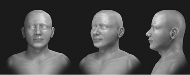
Fig. 8. Objective facial approximation rendering

Fig. 9. Complete – Three-quarter view

Fig. 10. Complete – Frontal and profile views
This work was the first proposed case of purely digital and three-dimensional facial approximation of the alleged Mozart skull. It was possible to compare the results with previous approximations, which showed convergence, serving as an illustrative case of structural coherence in forensic facial approximation, regrardless of applying approaches based on different techniques introduced in different decades. The use of artificial intelligence, performed with human monitoring and manual adjustments, allowed a significant increase in details, without clashing with the raw renderings, demonstrating that the use of these tools, instead of distorting the work of an expert, can be an important aid in improving graphic quality. Because it is an approach focused on the use of open source software, aiming at educational purposes for the field of forensic facial reconstruction, this work also served as a source of data for the replication of techniques, by sharing not only the step-by-step process, but also teaching tools that, together, allow the replication of the process by potentially interested parties.
Acknowledgements
To Dr. Richard Gravalos, for providing the CT scan used in this study.
Conflict of interests
The authors have no conflict of interests.
Authors’ contributions
CM: conceptualization, writing-first draft and revision; JŠ, ME.H, LS, TB: critical review of the first draft, writing-revision, literature search, methodology; EV, FMG: writing-first draft and revision, supervision.
Abdullah JY, Moraes C, Saidin M, Rajion ZA, Hadi H, Shahidan S, Abdullah JM. 2022. Forensic Facial Approximation of 5000-Year-Old Female Skull from Shell Midden in Guar Kepah, Malaysia. Appl Sci 12(15):7871. https://doi.org/10.3390/app12157871
Baldasso RP, Moraes C, Gallardo E, Stumvoll MB, Crespo KC, Strapasson RAP, de Oliveira RN. 2020. 3D forensic facial approximation: Implementation protocol in a forensic activity. J Forensic Sci 66(1):383–388. https://doi.org/10.1111/1556-4029.14587
Black A. 2012. Mozart’s Skull. Atlas Obscura. https://www.atlasobscura.com/places/university-mozarteum (accessed on October 4, 2024).
da Costa NR, Mancine L, Salvini R, Teixeira JM, Rodriguez RD, Leite REP, Nascimento C, Pasqualucci CA, Nitrini R, Jacob-Filho W, Lafer B, Grinberg LT, Suemoto CK, Nunes PV. 2022. Microcephaly measurement in adults and its association with clinical variables. Rev Saude Publica 56:38. https://doi.org/10.11606/s1518-8787.2022056004175
De Greef S, Claes P, Vandermeulen D, Mollemans W, Suetens P, Willems G. 2006. Large-scale in-vivo Caucasian facial soft tissue thickness database for craniofacial reconstruction. Forensic Sci Int 159: S126–146. https://doi.org/10.1016/j.forsciint.2006.02.034
Eng JE. 1906. Neuerlicher Zuwachs im Wohnzimmer der Familie Mozart. In: Mozarts Geburtshaus zu Salzburg, editor. Katalog des Mozart-Museums im Geburts- und Wohnzimmer Mozart’s zu Salzburg. Salzburg: Joh. Ev. Engl. 49–56.
Harding L. 2006. DNA detectives discover more skeletons in Mozart family closet. The Guardian. Online at: https://www.theguardian.com/world/2006/jan/09/arts.music (accessed on October 4, 2024).
Karhausen LR. 2001. The Mozarteum’s skull: a historical saga. J Med Biogr 9(2):109-117. https://doi.org/10.1177/096777200100900211
Kritscher H, Szilvássy J, Vlček E, Hauser G, Poxleitner-Blasl H, Sekal C. 1989. Zur Identifizierung des Mozartschädels. Ann Naturhist Mus Serie A 93:1–139.
Moraes C, Gravalos R, Machado CR, Chilvarquer I, Curi J, Beaini TL. 2022. Investigação de Preditores Anatômicos para o Posicionamento dos Globos Oculares, Asas Nasais, Projeção dos Lábios e Outros a partir da Estrutura do Crânio. Unpublished pre-print. figshare. https://doi.org/10.6084/M9.FIGSHARE.19686294
Moraes C, Sobral DS, Mamede A, Beaini TL. 2021. Sistema Complementar de Projeção Nasal em Reconstruções/Aproximações Faciais Forenses. Unpublished pre-print. figshare. https://doi.org/10.6084/M9.FIGSHARE.17209379
Moraes C, Dornelles R, Rosa ED. 2021. Sistema de Reconstrução de Tomografia Computadorizada Baseado no Slicer 3D e no DicomToMesh. Unpublished pre-print. figshare. https://doi.org/10.6084/M9.FIGSHARE.13513890
Moraes C, Suharschi I. 2022. Mensuração de Dados Faciais Ortográficos em Moldavos e Comparação com Outras Populações. Unpublished pre-print. https://doi.org/10.6084/m9.figshare.20089754.v1
Moraes C, Habicht ME, Galassi FM, Varotto E, Beaini T. 2023. Pharaoh Tutankhamun: a novel 3D digital facial approximation. Ital J Anat Embryol 127(1):13–22. https://doi.org/10.36253/ijae-14514
Moraes C, Beaini TL. 2024. A Aproximação Facial do Controverso Crânio Atribuído a Nicolau Copérnico (1473–1543). Unpublished pre-print: figshare. 10.6084/M9.FIGSHARE.25447210.
Moraes C, Galassi FM, Sineo L, Šindelář J, Varotto E, Mietlińska-Sauter J, Antunes-Ferreira N, Habicht ME, Beaini T. (2024). The Anatomical Bases of the 3D Digital Facial Approximation of the Zlatý kůň 1 Woman (ca. 43,000 BP). Anthropol Rev 87(2):85–97. https://doi.org/10.18778/1898-6773.87.2.04
Murray TJ. 1993. The skull of Mozart. Dalhousie Rev 73:246–251.
Pereira JGD, Magalhães LV, Costa PB, da Silva RHA. 2017. Reconstrução Facial Forense Tridimensional: Técnica Manual vs. Técnica Digital. Legal Rev Bras Odontol Leg 4(2):46–54. Legal. https://doi.org/10.21117/rbol.v4i2.111
Pinto RR, Almeida SMD, Chaves RBDN, Carvalho OAD, Machado MPS, Moraes C. 2020. Reconstrução Facial Forense de um Crânio Arqueológico com o ForensicOnBlender. Unpublished preprint. figshare. https://doi.org/10.6084/M9.FIGSHARE.12943418
Puech PF., Puech B, Tichy G, Cianfarani F, Albertini H. 1987. Les Dents de Wolfgang Amadeus Mozart. Bull Mém Soc Anthropol 4–3:207–212.
Puech PF, Puech B, Tichy G. 1989a. Identification of the cranium of W.A. Mozart. Forensic Sci Int 41(1–2):101–110. https://doi.org/10.1016/0379-0738(89)90241-7
Puech B, Puech PF, Tichy G, Dhellemmes P, Cianfarani F. 1989b. Craniofacial dysmorphism in Mozart’s skull. J Forensic Sci 34(2):487–490.
Puech B, Puech PF, Dhellemmes P, Pellerin P, Lepoutre F, Tichy G. 1989c. Did Mozart have a chronic extradural haematoma? Injury 20(6):327–330. https://doi.org/10.1016/0020-1383(89)90004-1
Puech PF. 1991. Forensic scientists uncovering Mozart. J R Soc Medicine 84(6):387. https://doi.org/10.1177/014107689108400646
Quatrehomme G, Cotin S, Subsol G, Delingette H, Garidel Y, Grévin G, Fidrich M, Bailet P, Ollier A. 1997. A fully three-dimensional method for facial reconstruction based on deformable models. J Forensic Sci 42(4):649–652.
Ritchie SJ, Cox SR, Shen X, Lombardo MV, Reus LM, Alloza C, et al. 2018. Sex Differences in the Adult Human Brain: Evidence from 5216 UK Biobank Participants. Cereb Cortex 28(8):2959–2975. https://doi.org/10.1093/cercor/bhy109
Sadie S. 2024. Wolfgang Amadeus Mozart. Encyclopædia Britannica. https://www.britannica.com/biography/Wolfgang-Amadeus-Mozart (Accessed on October 4, 2024).
Stephan CN. 2015. Facial Approximation-From Facial Reconstruction Synonym to Face Prediction Paradigm. J Forensic Sci 60(3):566–571. https://doi.org/10.1111/1556-4029.12732
Taylor KT. 2000. Forensic Art and Illustration. CRC Press. https://doi.org/10.1201/9781420036954
Wilkinson C. 2004. The Manchester Method of Facial Reconstruction. Forensic Facial Reconstruction. Cambridge: Cambridge University Press. https://doi.org/10.1017/cbo9781107340961.008

