Available online at: https://doi.org/10.18778/1898-6773.87.2.04
 https://orcid.org/0000-0002-9479-0028
Arc-Team Brazil, Sinop-MT, Brazil
 https://orcid.org/0000-0001-8902-3142
Department of Anthropology, Faculty of Biology and Environmental Protection, University of Lodz, Lodz, Poland
 https://orcid.org/0000-0001-8634-2295
Department of Biological, Chemical and Pharmaceutical Sciences and Technologies (STEBICEF), University of Palermo, Palermo, Italy
Department of Cultures and Societies, University of Palermo, Palermo, Italy
Surveyor, GEO-CZ, Tábor-Czech Republic
 https://orcid.org/0000-0001-6637-9402
Department of Cultures and Societies, University of Palermo, Palermo, Italy
Faculty of Dentistry, Federal University of Uberlândia, Uberlândia-MG, Brazil
 https://orcid.org/0000-0002-9185-7493
Department of Anthropology, Faculty of Biology and Environmental Protection, University of Lodz, Lodz, Poland
 https://orcid.org/0000-0002-0146-6369
Egas Moniz Center for Interdisciplinarity Research, Egas Moniz School of Health and Science, Portugal
 https://orcid.org/0000-0002-0193-9672
Archaeology, College of Humanities, Art and Social Sciences, Flinders University, Adelaide-SA, Australia
 https://orcid.org/0000-0003-0034-624X
Faculty of Dentistry, Federal University of Uberlândia, Uberlândia-MG, Brazil
ABSTRACT: In 1950 on Mount Zlatý kůň (‘Golden Horse’) in modern-day Czech Republic a system of caves was discovered. During many years of research in this area, human and animal osteological remains have been excavated, among which the most interesting ones were nine fragments of a female skull, now dated to ca. 43,000 yrs BP which are one of the earliest known anatomically modern humans in Eurasia. The aim of this research was to use purely digital techniques to: (1) to reconstruct the skull based on the 3D data of preserved fragments, (2) to approximate the probable appearance of the female it belonged to, and (3) to analyze the calculated shape of the reconstructed mandible and volume of the neurocranium in the context of similarities and differences with other representatives of the genus Homo. Computer techniques used in this research constitute a new, original approach to the problem of 3D analyses and may be useful primarily in bioarchaeological sciences, where metric analyses of the most valuable bone artifacts are often severely limited due to the incompleteness of the material available for research. The digital techniques presented here may also contribute significantly to the field of surgery, with the possibility of being adapted for applications in cranial prosthetics and post-traumatic reconstructive surgery.
KEY WORDS: Zlatý kůň 1; facial approximation; digital; anatomy; prehistory; anthropology.
In 1950, during the works on the explosion of a large limestone rock on Mount Zlatý kůň (‘Golden Horse’), the Koněprusy cave system (in modern-day Czech Republic) was discovered by construction workers. Its exploration over the following years revealed the presence of human and animal remains, as well as stone- and bone-made artifacts attributed to the early Upper Palaeolithic period. Attention was paid to the presence of what initially appeared to be two separate skulls, but later, when the pieces of bone were assembled, were argued to belong to a single individual. Contradictory studies continued to emerge, as, given the structural robustness, the remains were firstly attributed to a male although subsequent analyses indicated an adult female. A similar problem arose in the case of assessing the antiquity of the remains: initially, estimated to be ca. 30,000 years old by the archaeological stratigraphic method and then, using radiocarbon dating, reconsidered to be much younger, ca. 15,000 before present (BP). However, a craniometric analysis found the remains compatible with a chronology anterior to the Last Glacial Maximum (LGM, ca. 20,000 years ago). Later 14C tests dated the remains back to ca. 34,000 years BP, but it was believed that the skeleton was too contaminated by an animal glue found on the skeletal elements to yield a reliable absolute dating. Genomic analyses carried out on the remains and Bayesian tip dating suggested a much earlier chronology of ca. 43,000 years ago, making the Zlatý kůň 1 woman one of the earliest known anatomically modern humans preserved from the stock of the first Eurasian inhabitants (Posth 2021; Prüfer et al. 2021; Rmoutilová et al. 2018; Svoboda 2000). Palaeogenetically, this female individual’s genome was shown to have a limited amount of Neanderthal admixture and it is worth underlining how the lineage she belonged to did not contribute genetically to later generations of people in Europe or Asia (Churchill et al. 2022; Prüfer et al. 2021). In this article, a 3D facial approximation based on an anatomical analysis is offered using novel digital techniques.
Forensic facial reconstruction (FFR) or forensic facial approximation (FFA) (Stephan 2015) is an auxiliary recognition technique that predicts facial appearance and is used when little information is available to identify an individual based on their remains (Pereira et al. 2017). It should be stressed that this technique is not about making an exact identification, such as those provided by DNA tests or comparative analysis of teeth, but, rather, it relies on the recognition of the facial aspects that may indirectly lead to individual’s identification. As previously noted, for the FFA process to be feasible, it is necessary to first obtain the appearance of the skull itself.
This skull image was acquired in a purely digital process, based on previously published methodology, an approach which will be described in further detail later. Facial approximation followed the same approach described in Abdullah et al. (2022) and Moraes (2023), although with small modifications. The modeling process was carried out in the Blender 3D software, running the add-on OrtogOnBlender (http://www.ciceromoraes.com.br/doc/pt_br/OrtogOnBlender/index.html) and its submodule ForensicOnBlender. The program and add-on are free, open source and multiplatform. They can run on Windows (>=10), MacOS (>=BigSur) and Linux (=Ubuntu 20.04).
In the present work, a desktop computer with the following characteristics was used:
like in the list above Linux 3DCS (https://github.com/cogitas3d/Linux3DCS), based on Ubuntu 20.04.
The skull known as Zlatý kůň 1 consists of nine fragments (codes: AP2, AP3, AP9, AP10, AP12, AP15, AP18, AP18 and AP21), which are stored at the Anthropology Department of the National Museum, located in Prague, Czech Republic. Despite covering a considerable part of the surface of a composite skull (cranium and mandible), the structure has some missing regions, such as the nasal bone, part of the maxilla, the left orbit, and the left part of the frontal bone. In 2018, a multinational team of researchers carried out the work of three-dimensional reconstruction of the missing regions, using statistical data extracted from a group made up of 31 skulls: 30 modern ones (15 males and 15 females, scanned using computed tomography at the Center Hospitalier Universitaire, CHU, in Bordeaux, France) and one of the Moča skull, found in Slovakia, dated to 13,100 years BP. The researchers initially mirrored the 3D mesh to reconstruct the missing regions, using the original anatomy of the skull. Since the image obtained using this method still had some empty areas, it was then complemented basing on statistical data extracted from the aforementioned tomography and fossils, which led to obtaining an image of a complete skull (Rmoutilová et al. 2018). Unfortunately, the authors of this study did not have direct access to the Zlatý kůň 1 fossils, therefore they decided to reconstruct the skull based on data available in scientific publications (Posth 2021; Prüfer et al. 2021; Rmoutilová et al. 2018; Svoboda 2000).
This study uses the approach of Moraes et al. (2023), previously used, among others, in the reconstruction of the skull of Pharaoh Tutankhamun. The facial approximation discussed in this research used as a reference the skull reconstructed by Rmoutilová et al. (2018), based on the images available in the publication (open access under the Creative Commons license) in order to deform the skull of a virtual donor over the spatial references, thus correcting the structure with the measurement data present in the same material (Rmoutilová et al. 2018), and reinforcing the precision of the scale with data collected from Prüfer et al. (2021). The anatomical deformation resulted in a skull closely approximating the fossil Zlatý kůň 1 (Fig. 1A). At first, the structure of the zygomatic arch, close to the porium, seemed to differ from the expected anatomical pattern. Aiming to compare with modern individuals, a series of 30 skulls of different population affinities and sexes received a two-dimensional tracing with orthographic observation along the X axis in order to establish a lateral pattern of the region (Fig. 1 B). Two other fossils received the same graphic treatment and, at the end, the set of 30 modern samples (in gray) were compared to the fossils Zlatý kůň 1 (in green), Mladeč 1 (in red, Moraes et al. 2022) and Nazlet Khater 2 (in blue, Moraes and Santos 2023). This comparison indicates a similar initial elevation of the zygomatic arch in the three fossils, which clearly differs from the group of modern skulls (Fig. 1C). With the issue related to the zygomatic arch overcome, attention turned to other regions, with the projection of lines and limits expected for the skull and soft tissue (Moraes et al. 2022a; Moraes and Suharschi 2022; two classes on this approach are available online, 1 of 2: https://youtu.be/U6oYkEmfyWo, 2 of 2: https://youtu.be/Vcz2e5uSFX8). The projection of the lines was compatible with the skull reconstructed by Rmoutilová et al. (2018), except the orbital frontomalar distance (fmo-fmo) and the projection of other measurements based on the fmo-fmo ratio. According to the fmo-fmo proportion, the limit of the incisors would be below the reconstructed skull, as well as below the chin (Fig. 1D, in blue). However, when taking into account the expected mean for such regions, the lines are close and within one standard deviation (Fig. 1E). The difference between the fmo-fmo ratio and the average can be explained by the distance between the gonions, generally compatible with the fmo-fmo distance, but which, in this case, was significantly smaller. While the fmo-fmo distance was ca. 102 mm, compared to the general average of ca. 97 mm (https://bit.ly/3NRw2KW), the go-go distance was ~94 mm, compared to an average of ca. 97 mm. Therefore, the mandible proportion is more appropriate to the average than the proportion expected by the fmo-fmo distance. The data reinforces the statistical coherence of the reconstruction carried out by the multinational team in 2018 (Rmoutilová et al. 2018).

Fig. 1. Three-dimensional reconstruction of the skull
Since the human skull discussed in this work has poorly understood context of population affinities (apparently the population to which this individual belonged did not genetically contribute to either Europeans or modern Asians, Prüfer et al. 2021), this work disregarded the use of thickness markers of soft tissues and the authors chose to use only the anatomical deformation on the fossil. This approach proved to be very compatible with the parameters coming from the soft tissue thickness tables, keeping the limits within the SD in other approaches that used the technique (vd. OrtogOnLineMag #5: https://ortogonline.com/doc/pt_br/OrtogOnLineMag/5/ and OrtogOn-LineMag #6: https://ortogonline.com/doc/pt_br/OrtogOnLineMag/6/).. To reinforce volumetric precision, two 3D meshes from virtual donors were imported, including the skull, endocranium and soft tissue of a man and a woman, both adults (class available on anatomical deformation: https://youtube.com/xig5_EcIFWA). The deformed meshes received a line indicating the profile of the face, interpolating the two limits of the skin resulting from the anatomical deformation (Fig. 2 A, B), which were compatible with the nasal projection based on statistical data collected in computed tomography scans of living individuals belonging to different population affinities (Moraes et al. 2021; Moraes and Suharschi 2022). The limits projected from the skull were compared with the deformed mesh and adapted to the expected parameters, including the size of the nose on the X axis, the size of the eyes on the X axis, the position of the eyeball on the X, Y and Z axes, the size of the ears on the Z axis and the size of the lips on the X axis (Fig. 2C). Thanks to the pre-segmented structure, it was possible to adjust the mesh so that it resulted in the volume of the endocranium, whose data will be detailed later (Fig. 2D). Following the approach exposed in Abdullah et al. (2022) and Moraes (2023), a bust from another facial approximation was imported and adjusted to provide a mesh composed of four-sided faces and with a previously configured texture (Fig. 2E). Unlike the approaches mentioned above where a version with eyes open was available, the present work only has a version with eyes closed, in order to reduce the subjective elements of the structure, as will be explained in the Results. The mesh underwent detailing via digital sculpting, texture adjustment and scene lighting configuration so that the final images could be generated (Fig. 2. F, G).

Fig. 2. Steps of the digital facial approximation
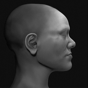
The final images were generated using Blender 3D’s Cycles renderer (https://www.blender.org/) and consisted of views of the bust composed to show the most objective elements of the face, focusing on the general structure. For the images with objective elements, the eyes were closed, the image was converted to gray scale, and the head did not have hair (Figs. 3–6). For images with speculative elements, the eyes were opened, the hair was configured, and the colors were maintained (Figs. 7–9).

Fig. 3. 3/4 image of the face approximated with objective elements

Fig. 4. Side image of the face approximated with objective elements

Fig. 5. Profile image of the approximate face with objective elements

Fig. 6. Frontal image of the face approximated with objective elements

Fig. 7. 3/4 image of the face approximated with speculative elements

Fig. 8. Side image of the face approximated with speculative elements

Fig. 9. Frontal image of the face approximated with speculative elements
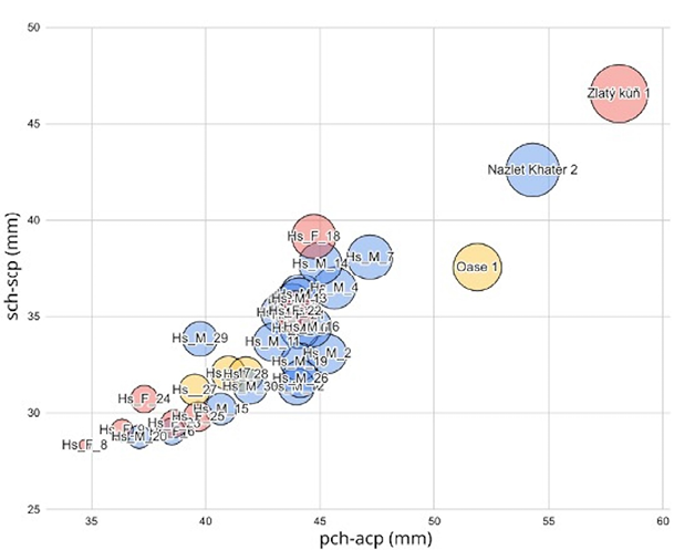
The 3D approximation of the examined specimen, obtained according to the adopted methodologies, makes it possible to make inferences on the morphology of the mandible and the endocranium volume between different species of Homo. An attempt to understand the differences between the mandibles of fossils dating from 32,000 to 45,000 BP, the bones of 30 modern individuals and the fossils Zlatý kůň 1, Oase 1 (Crevecoeur 2012) and Nazlet Khater 2 (Moraes and Santos 2023) were measured at two distances, sch-scp and pch-acp (Fig. 10), both only on the Y axis, with sch being point on superior margin of the condyle head, scp – point on superior margin of the coronoid process, pch – point on the posterior margin of the condyle head and acp – most anterior point on the anterior aspect of the ramus border (Lestrel et al. 2013). A graph (Fig. 11) created on the base of pch-acp measurements compares three fossils: Oase 1 (approx. 40,000 years ago), Nazlet Khater 2 (32,000–44,000 years ago) and Zlatý kůň 1 (approx. 43,000 years ago), as well as broad group of modern human mandibles. As can be seen, Zlatý kůň 1 is characterised by the significantly most robust mandible structure. By adding data for four mandibles belonging, according to Rosas et al. 2019, to H. neanderthalensis (Atapuerca-605, Atapuerca-905, Mauer and Arago 2), three distinct groups can be created (Fig. 12). First, a broader one, consists of the H. neanderthalensis individuals, which slightly intersects with second group – modern humans. This group is characterised by the smallest mandibles. The third group is formed by H. sapiens, which comprises fossils aged from 32,000 to 45,000 yrs BP. It is clearly visible that this group is distant from modern humans but intersects with H. neanderthalensis. Something similar happens with the data related to the endocranium volume (X axis) and head circumference (Y axis, see Fig. 13). Points representing fossils of individuals living between 31,000 and 45,000 yrs BP, for the most part, touch the ellipse composed of H. neanderthalensis, H. rhodesiensis and H. heidelbergensis. The volume of Zlatý kůň 1’s endocranium resulted in ca.1590 cm³, a value above one SD from the average of current female. In relation to the head circumference, with 59.08 cm, also above one SD from the average.

Fig. 10. Measurements taken on the mandible (Y axis)

Fig. 11. Distribution of pch-acp measurements on the X axis and sch-scp on the Y axis, the diameter of the spheres is proportional to the sum of the two measurements (pch-acp)+(sch-scp). The colors represent sex, with male blue, female red and undefined yellow

Fig. 12. Distribution of pch-acp measurements on the X axis and sch-scp on the Y axis, with the addition of the H. neanderthalensis group
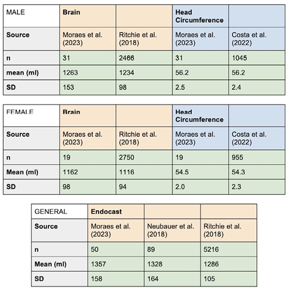
The endocranium volume and circumference graph was generated based on data for a group of 50 H. sapiens endocrania, 31 male and 19 female (Fig. 13). When comparing this data to outcomes based on noticeably larger numbers (up to 2750 for each sex, da Costa et al. 2022; Neubauer et al. 2018; Ritchie et al. 2018), similar sample’s distribution is still visible (Fig. 14). When applying the factor of -9.81% to convert the volume of the Zlatý kůň 1 endocranium into brain volume (Moraes et al. 2023), 1434 cm³ is reached, that is, 318 cm³ above the average, which is 1116 cm³. Therefore, when using data from Ritchie et al. (2018), Zlatý kůň 1’s brain is 3.53 SD above the average for females. Even if compared to males, the brain would be 2.0 SD above the average, which is 1234 cm³. The head circumference, which in Zlatý kůň 1 individual resulted in 59.08 cm, is 2.08 SD above the female average according to Costa et al. (2022). When taking into account the group of both sexes of H. sapiens, the endocranium (not the brain) of Zlatý kůň 1 is also 1.6 SD above the general average, according to Neubauer et al. (2018). The volumetric and linear data measured on the fossil Zlatý kůň 1 are based on the information provided by Prüfer et al. (2021) and Rmoutilová et al. (2018). Zlatý kůň 1’s encephalic size is compatible with evolutionary trends affecting anatomically modern H. sapiens in that during the Holocene period (last 10,000 years, hence much later than the time in which this individual lived) brain size shrunk by approximately 10% (Ruff et al. 1997; Henneberg 1988; Neubauer et al. 2018).

Fig. 13. Endocranium volume vs head circumference

Fig. 14. Comparison between different studies
The rapidly developing visualisation and measurement techniques using 3D models are increasingly applied not only in engineering sciences, but also in medical ones, especially surgery, and scientific research, including physical anthropology. This study aimed to present a new, original method used in morphometric analyzes of bone remains and approximation of the real appearance of a face of the Zlatý kůň 1 woman, one of the earliest known Eurasian individuals. Due to the lack of access to the original remains, this study was based purely on digital data published in the scientific press. Based on digital data, the spatial arrangement of the preserved bone fragments was determined, and then the missing elements were reconstructed, ultimately obtaining an image of a complete skull and an approximation of the probable life appearance of the woman it belonged to. The obtained results were compared to those published in previous works that dealt with the reconstruction of the appearance of the Zlatý kůň 1 woman. Finally, the shape of the mandible reconstructed using digital methods and the volumes of the endocranium (1590 cm³) and brain (1434 cm3) were discussed, and then a comparison was made with currently known representatives of other species of the genus Homo. The calculated dimensions place the examined individual in the group of other examples of H. sapiens living between 45,000–32,000 years BP, which is consistent with the C14 dating of the material performed by independent researchers (43,000 years BP) and compatible with the described evolutionary trends of decreasing brain dimensions and decline massiveness of the mandible of our species. These studies show that the use of modern research methods can significantly increase knowledge in the field of morphometric research, which is particularly valuable in the case of often fragmentarily preserved bone remains representing the ancestors of modern humans.
Acknowledgements
The latest version of open-access pre-print of an earlier version of this manuscript was posted in an online repository at https://doi.org/10.6084/m9.figshare.23733504 on July 18, 2023. This pre-print was referenced by journalists covering the story for the general public, e.g. https://www.livescience.com/archaeology/see-stunning-likeness-of-zlaty-kun-the-oldest-modern-human-to-be-genetically-sequenced. Nonetheless, the present re-worked version represents the finalized scientific product submitted for peer-review and potential scientific publication. The authors would like to thank Dr. Richard Gravalos for providing the CT scans of the virtual donors used in this study. To Lis Moura, for the important contribution in suggesting the comparison of the zygomatic structure with other fossils from a similar period. Moreover, the authors would like to specify that the Creative Commons license Attribution 4.0 International (CC BY 4.0) has been followed for the graphic material used in this manuscript.
Funding
No funding was received for conducting this study.
Conflict of interests
None to disclose.
Authors' contribution
CM: conceptualisation, analysis, 3D modelling, writing of the first draft; FMG: conceptualisation, analysis, writing of the first draft, literature search; LS: analysis, literature search, critical revision of the first draft; JŠ: analysis, literature search, critical revision of the first draft; EV: conceptualisation, analysis, writing of the first draft, literature search; JM-S: analysis, literature search, critical revision of the first draft; NAF: analysis, literature search, critical revision of the first draft; MEH: analysis, literature search, critical revision of the first draft; TB: analysis, literature search, critical revision of the first draft, supervision.
Abdullah JY, Moraes C, Saidin M, Rajion ZA, Hadi H, Shahidan S, Abdullah JM. 2022. Forensic Facial Approximation of 5000–Year-Old Female Skull from Shell Midden in Guar Kepah, Malaysia. Applied Sciences (Switzerland) 12. https://doi.org/10.3390/app12157871
Churchill SE, Keys K, Ross AH. 2022. Midfacial Morphology and Neandertal–Modern Human Interbreeding. Biology 11:1163. https://doi.org/10.3390/biology11081163
da Costa NR et al. 2022. Microcephaly measurement in adults and its association with clinical variables. Revista de Saude Publica 56: 1–10. https://doi.org/10.11606/s1518-8787.2022056004175
Crevecoeur I. 2012. The Upper Paleolithic Human Remains of Nazlet Khater 2 (Egypt) and Past Modern Human Diversity. Vertebrate Paleobiology and Paleoanthropology 2:205–219. https://doi.org/10.1007/978-94-007-2929-2_14
Henneberg M. 1988. Decrease of human skull size in the Holocene. Hum Biol 60:395–405.
Lestrel PE, Wolfe CA, Bodt A. 2013. Mandibular shape analysis in fossil hominins: Fourier descriptors in norma lateralis. HOMO- Journal of Comparative Human Biology 64:247–272. https://doi.org/10.1016/j.jchb.2013.05.001
Moraes C. 2023. A Aproximação Facial Digital 3D de Ava (Escócia, ~3806 AP). https://doi.org/10.6084/m9.figshare.23560587
Moraes C, Gravalos R, Machado CR, Chilvarquer I, Curi J, Beaini TL. 2022a. Investigação de Preditores Anatômicos para o Posicionamento dos Globos Oculares, Asas Nasais, Projeção dos Lábios e Outros a partir da Estrutura do Crânio. In: OrtogOnLineMag #4, Moraes C (ed). 51–93. https://doi.org/10.6084/m9.figshare.19686294.v1
Moraes C, Habicht ME, Galassi FM, Varotto E, Beaini T. 2023. Pharaoh Tutankhamun: a novel 3D digital facial approximation. Italian Journal of Anatomy and Embryology 127:13–22. https://doi.org/10.36253/ijae-14514
Moraes C, Santos ME. 2023. A Aproximação Facial do Crânio de Nazlet Khater 2. In OrtogOnLineMag #6, Moraes C (ed). 7–15. https://doi.org/10.6084/m9.figshare.22557598
Moraes C, Šindelář J, Drbal K. 2022b. A Aproximação Facial Forense do Crânio Mladeč 1. In OrtogOnLineMag #5, Moraes C (ed). 1–27. https://doi.org/10.6084/m9.figshare.20435787
Moraes C, Sobral DS, Mamede A, Beaini TL. 2021. Sistema Complementar de Projeção Nasal em Reconstruções/Aproximações. In OrtogOnLineMag 3, Moraes C (ed). 37–54. https://doi.org/10.6084/m9.figshare.17209379.v1
Moraes C, Suharschi I. 2022. Mensuração de Dados Faciais Ortográ fi cos em Moldavos e Comparação com Outras Populações. In OrtogOnLineMag #4, Moraes C (ed). https://doi.org/10.6084/m9.figshare.20089754
Neubauer S, Hublin JJ, Gunz P. 2018. The evolution of modern human brain shape. Science Advances 4: 1–8. https://doi.org/10.1126/sciadv.aao5961
Pereira JGD, Magalhães LV, Costa PB, Silva RHA da. 2017. Reconstrução Facial Forense Tridimensional: Técnica Manual Vs. Técnica Digital. Revista Brasileira de Odontologia Legal 4: 46–54. https://doi.org/10.21117/rbol.v4i2.111
Posth C. 2021. A bumpy ride to the re-discovery of Zlatý Kůň. Nature News [online] Available at: https://communities.springernature.com/posts/a-bumpy-ride-to-the-re-discovery-of-zlaty-kun
Prüfer K, Posth C, Yu E, Stoessel A, Spyrou MA, Deviese T, et al. 2021. A genome sequence from a modern human skull over 45,000 years old from Zlatý kůň in Czechia. Nature Ecology and Evolution 5: 820–825. https://doi.org/10.1038/s41559-021-01443-x
Ritchie SJ, Cox SR, Shen X, Lombardo MV, Reus LM, Alloza C, et al. 2018. Sex differences in the adult human brain: Evidence from 5216 UK biobank participants. Cerebral Cortex 28: 2959–2975. https://doi.org/10.1093/cercor/bhy109
Rmoutilová R, Guyomarc’h P, Velemínský P, Šefčáková A, Samsel M, Santos F, Maureille B, Brůžek J. 2018. Virtual reconstruction of the upper palaeolithic skull from zlatý kůň, Czech Republic: Sex assessment and morphological affinity. PLoS ONE 13:1–29. https://doi.org/10.1371/journal.pone.0201431
Rosas A, Bastir M, Alarcón JA. 2019. Tempo and mode in the Neandertal evolutionary lineage: A structuralist approach to mandible variation. Quaternary Science Reviews 217:62–75. https://doi.org/10.1016/j.quascirev.2019.02.025
Ruff CB, Trinkaus E, Holliday TW. 1997. Body mass and encephalization in Pleistocene Homo. Nature 387: 173–176. https://doi.org/10.1038/387173a0
Stephan CN. 2015. Facial Approximation-From Facial Reconstruction Synonym to Face Prediction Paradigm. Journal of Forensic Sciences 60:566–571. https://doi.org/10.1111/1556-4029.12732
Svoboda J. 2000. The depositional context of the Early Upper Paleolithic human fossils from the Koneprusy (Zlatý kůň) and Mladec Caves, Czech Republic. Journal of Human Evolution 38: 523–536. https://doi.org/10.1006/jhev.1999.0361

