Comparative Analysis of Foot Arch Index between Yogic and Non-Yogic Female Practitioners
 https://orcid.org/0000-0003-1768-0631
Department of Physical Education, Faculty of Arts, Banaras Hindu University, Varanasi, India, 221005
School of Sports Studies, TransStadia University, Ahmedabad, India, 380022
https://orcid.org/0000-0001-8051-341X
Department of Physical Education, Faculty of Arts, Banaras Hindu University, Varanasi, India, 221005
https://orcid.org/0000-0002-0765-1944
Department of Physical Education, Faculty of Arts, Banaras Hindu University, Varanasi, India, 221005
Abstract
Introduction
With advancing age in females, gait patterns change due to several factors, including nutritional and hormonal influences. These changes can lead to a lowering of the foot arch, resulting in mild valgus and varus deformities. Regular yoga practice may help tone the musculoskeletal system of the foot, promoting the maintenance of the medial longitudinal, transverse, and lateral arches.
Study Aim
This cross-sectional study aimed to compare the arch indices of female yoga practitioners to those of non-practitioners in order to ascertain how yoga affected female arch indices.
Materials and Methods
Fourteen female students from Banaras Hindu University in Varanasi, India, participated in the study, seven of which were regular yoga practitioners and seven were non-yoga practitioners. Their left and right arch indices were determined by taking measurements from their footprints. A Mann Whitney U test was used to compare the foot arch indices of the two groups.
Results
A statistically significant difference between the left as well as right foot arch indices of females was revealed.
Conclusion
The study concludes that females who practice yoga may experience foot arch plasticity which is not necessarily evident in non-yoga practitioners. Future studies should test larger sample sizes and different genders.
Keywords: foot biomechanics, foot health, physical activity, exercise, body adaptation
Introduction
Importance of the Foot Arch and Its Role in Bipedal Locomotion
The human foot plays a vital role in supporting bipedal locomotion, with the medial longitudinal arch (MLA) serving as a key structural component in shock absorption, balance, and weight distribution. This arch allows the foot to adapt to uneven surfaces and absorb forces during gait. When the MLA is compromised, it can result in biomechanical dysfunctions, altered gait, and discomfort that may impair walking and posture (Holowka & Lieberman, 2018; Ker et al., 1987). The MLA is a unique feature in human evolution, enabling effective upright walking and running. Pathologies such as plantar fasciitis, Achilles tendinitis, and Morton’s neuroma are known to cause foot pain and may limit mobility. Similarly, deformities like pes planus (flat feet) and pes cavus (high arches) often develop from prolonged weight-bearing or overuse and can be associated with secondary issues, including low back pain (Tsung et al., 2003). These facts emphasize the need for regular assessment of MLA using reliable, non-invasive tools to ensure early detection and correction.
Tools for Assessing the Medial Longitudinal Arch
Several tools and indices have been developed to evaluate the structure and function of the MLA. Among these, the Foot Posture Index (FPI), and its revised version FPI-6, are widely used for visually assessing foot alignment in clinical and research settings. The FPI-6 has demonstrated moderate to high intra-rater reliability and moderate inter-rater reliability, and it has been validated in various age groups (Keenan et al., 2007; Morrison & Ferrari, 2009). In pediatric populations, the FPI-6 has shown good diagnostic accuracy when compared to radiographic assessments, with reported sensitivity and specificity values above 80% (Lee et al., 2020).
In addition to observational indices, footprint-based tools such as the Arch Index (AI) (Cavanagh & Rodgers, 1987) and the Staheli Index (Staheli et al., 1987) are frequently employed. These indices offer a non-invasive, cost-effective method for estimating MLA height. Although radiographic and anthropometric methods are more precise, footprint-based indices are widely used in field studies due to their simplicity. Importantly, Kanatli et al. (2001) reported significant correlations between footprint indices and radiographic measures of MLA, supporting their clinical relevance. Despite some limitations in accuracy, footprint indices remain a practical option, especially when radiological evaluation is not feasible.
Influence of Body Composition and Lifestyle Factors on Medial Longitudinal Arch
The height and structure of the MLA are influenced not only by mechanical factors but also by body composition, gender, age, and lifestyle. Obesity has been identified as a major risk factor for pes planus, where increased body mass may cause excessive flattening of the arch, compromising foot function and resilience (Butler et al., 2008). This arch flattening can also lead to secondary problems in the knees, hips and lower back.
Several studies have explored the relationship between MLA parameters and body mass index (BMI). Stanković et al. (2018) observed a strong correlation between left foot arch angle and BMI, suggesting that this angle may serve as a more reliable predictor of BMI compared to other indices. Gilmour and Burns (2001) emphasized that body composition significantly affects arch index values, highlighting the importance of interpreting such indices alongside body composition data. Other variables, including gender, limb dominance, and age, have also been shown to influence MLA structure. In a study by Wearing et al. (2004), both AI and Navicular Height (NH) were found to be valid, non-invasive predictors of MLA across age groups. While NH appeared more sensitive to age-related changes, AI was slightly more accurate overall. Together, these findings support the use of such indices in large-scale population studies.
In light of the growing interest in the role of physical activity in foot structure, this study investigates whether regular yoga practice has an effect on the MLA in females. We test a hypothesis that the foot arch index of female yoga practitioners will differ from non-practitioners.
Materials and Methods
A total of fourteen female participants were selected through simple random sampling from various sports specializations at the Department of Physical Education, Banaras Hindu University, India, aged between 19–23 years. Among them, seven participants reported engaging in regular yoga practice, while the remaining seven had no prior experience with yoga or related physical activities. Prior to data collection, all participants received both verbal and practical orientation regarding the purpose and procedures of the study. The study involved non-invasive procedures limited to the collection and analysis of static footprints. All participants were healthy adult volunteers who were informed in detail about the aims, procedures, and voluntary nature of the research. Written informed consent was obtained from each participant prior to data collection. As the study did not involve any clinical intervention, collection of sensitive personal data, or work with vulnerable populations, formal ethical clearance was not required by our institution. Nonetheless, the study adhered to the ethical principles outlined in the Declaration of Helsinki and followed standard academic practices for research involving human participants.
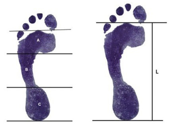
The primary variable selected for this study was the Foot Arch Index (Cavanagh & Rodgers, 1987; Ozer & Barut, 2012) (Figure 1), assessed separately for the left and right foot of each participant. This variable was chosen to evaluate the structure and characteristics of the MLA across both yogic and non-yogic groups. The Arch Index is the ratio of the area of the middle third of the toeless footprint to the overall toeless footprint area. A line is drawn between the center point of the second toe and the posterior-most point on the heel. Two parallel lines perpendicular to this line are drawn to divide the toeless footprint area into equal thirds. This was calculated from the foot imprints of the subjects, which were taken by using an inkpad on an A4 sheet with one leg standing position (Ozer & Barut, 2012).
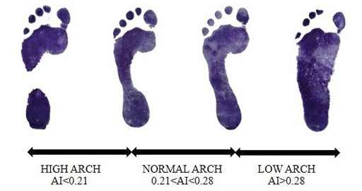
The Foot Arch Index was calculated based on the method described by Ozer and Barut (2012), which has been established as a reliable and valid approach for assessing foot morphology in both athletic and general populations. This method enables a consistent and non-invasive measurement of the medial longitudinal arch using static footprint data. To classify the arch types into high arch, normal arch, and low arch (pes planus), categorization thresholds were adopted from established normative data presented in previous research (Menz et al., 2012). These classification criteria were used to interpret individual Foot Arch Index values and group participants accordingly. The procedure and classification system are illustrated in Figure 2.
Statistical Procedures
Descriptive statistics, including the mean and standard deviation, were computed to summarize the arch index values of both yoga practitioners and non-practitioners. Given the small sample size (n = 14) and the non-parametric nature of the data, a Mann-Whitney U test was employed to examine differences in foot arch index between the two independent groups. All statistical analyses were performed using standard statistical software, with the level of significance set at p < 0.05.
Results
The mean and standard deviation of the left and right foot arch index for both yoga practitioners and non-yoga practitioners are shown in Table 1. The data indicate a lower average arch index in the yoga group, suggesting a comparatively higher medial longitudinal arch.
| Group | N | Mean Arch Index |
Standard Deviation |
|---|---|---|---|
| Yoga Practitioners: left foot | 7 | 0.264 | 0.052 |
| Non-Yoga Practitioners: left foot | 7 | 0.370 | 0.036 |
| Yoga Practitioners: right foot | 7 | 0.276 | 0.053 |
| Non-Yoga Practitioners: right foot | 7 | 0.360 | 0.037 |
Statistically significant differences were observed between the yoga practicing and non-practicing groups, indicating a potential influence of yoga practice on foot arch morphology. The p value was lower when the left foot was compared (U = 1; p=0.001), whereas it was higher (U =3 ; p=0.004), but still statistically significant when comparing the right foot.
Discussion
Our preliminary findings suggest that regular engagement in yoga may be associated with measurable alterations in medial longitudinal arch structure. The human foot, particularly the MLA, has long been considered an essential structure in the context of bipedal locomotion and postural stability—key traits in human evolutionary adaptation. This study explored whether regular engagement in yoga, a culturally rooted and functionally diverse physical practice, contributes to measurable differences in foot arch structure among females. The findings revealed statistically significant differences in the arch index of both the left and right foot between yoga practitioners and non-practitioners, suggesting that sustained functional loading through yoga may lead to subtle morphological adaptations in the foot.
From an anthropological perspective, these results add to the growing body of research that underscores the dynamic interaction between physical activity, cultural behavior, and human form. The consistent practice of yoga, involving postures that load the feet in varied planes and require proprioceptive feedback, may stimulate musculoskeletal adaptations over time—supporting the hypothesis that lifestyle factors can influence phenotypic variation in foot morphology (Kulthanan et al., 2004).
While previous research has emphasized the reliability of arch height index and footprint-based analysis in assessing MLA structure (McPoil et al., 2006; Ozer & Barut, 2012), the present findings highlight yoga as a potential behavioral modifier of foot form. The use of the arch index as a non-invasive measure aligns well with anthropological methods focused on external morphology and allows for cost-effective, reproducible data collection in field and community-based research.
The observed differences in arch index may reflect adaptive responses of the intrinsic and extrinsic foot musculature, possibly supporting arch elevation and alignment. These adaptations could reduce the risk of common foot pathologies such as pes planus, particularly in females, who are often more susceptible due to footwear choices and occupational load (Barton et al., 2009). Such findings are relevant in evolutionary anthropology and anatomy, where foot structure is not only examined for its role in past mobility patterns but also as a site of modern functional adaptation (e.g., Huang et al., 2019; Shibuya et al., 2010; Willems et al., 2012).
Nonetheless, this study is limited by its small, gender-specific sample size and cross-sectional design. Future studies should incorporate a broader demographic spectrum, including both sexes and varying age groups, to explore whether similar adaptations occur across populations. Longitudinal studies would also help determine whether observed changes are transient or structural.
Conclusion
The present study provides preliminary evidence that regular yoga practice may influence the medial longitudinal arch structure in women, as reflected in lower arch index values compared to non-practitioners. These differences suggest that cultural practices like yoga—when performed consistently—can serve as functional stimuli capable of inducing measurable morphological variation in the human foot. With further evidence from other studies, implications for both clinical health and anthropological research can be achieved. Continued investigation into the effects of habitual activity patterns on skeletal and soft tissue structures may further illuminate the plasticity of human form in response to lifestyle, and deepen our understanding of the role behavior plays in shaping the body across both evolutionary and contemporary time scales.
References
Barton, C. J., Bonanno, D., & Menz, H. B. (2009). Development and evaluation of a tool for the assessment of footwear characteristics. Journal of Foot and Ankle Research, 2(1), 10. https://doi.org/10.1186/1757-1146-2-10
Butler, R. J., Hillstrom, H., Song, J., Richards, C. J., & Davis, I. S. (2008). Arch height index measurement system: establishment of reliability and normative values. Journal of the American Podiatric Medical Association, 98(2), 102–106. https://doi.org/10.7547/0980102
Cavanagh, P. R., & Rodgers, M. M. (1987). The arch index: a useful measure from footprints. Journal of Biomechanics, 20(5), 547–551. https://doi.org/10.1016/0021-9290(87)90255-7
Gilmour, J. C., & Burns, Y. (2001). The measurement of the medial longitudinal arch in children. Foot & Ankle International, 22(6), 493–498. https://doi.org/10.1177/107110070102200607
Holowka, N. B., & Lieberman, D. E. (2018). Rethinking the evolution of the human foot: insights from experimental research. Journal of Experimental Biology, 221(17), jeb174425. https://doi.org/10.1242/jeb.174425
Huang, P., Liang, M., & Ren, F. (2019). Assessment of long-term badminton experience on foot posture index and plantar pressure distribution. Applied Bionics and Biomechanics, 2019(1), 8082967. https://doi.org/10.1155/2019/8082967
Kanatli, U., Yetkin, H., & Cila, E. (2001). Footprint and radiographic analysis of the feet. Journal of Pediatric Orthopaedics, 21(2), 225–228.
Keenan, A. M., Redmond, A. C., Horton, M., Conaghan, P. G., & Tennant, A. (2007). The Foot Posture Index: Rasch analysis of a novel, foot-specific outcome measure. Archives of Physical Medicine and Rehabilitation, 88(1), 88–93. https://doi.org/10.1016/j.apmr.2006.10.005
Ker, R. F., Bennett, M. B., Bibby, S. R., Kester, R. C., & Alexander, R. M. (1987). The spring in the arch of the human foot. Nature, 325(6100), 147–149. https://doi.org/10.1038/325147a0
Kulthanan, T., Techakampuch, S., & Donphongam, N. (2004). A study of footprints in athletes and non-athletic people. Journal-Medical Association of Thailand, 87(7), 788–793.
Lee, M. S., Vanore, J. V., Thomas, J. L., Catanzariti, A. R., Kogler, G., Kravitz, S. R., Miller, S.J., & Gassen, S. C. (2005). Diagnosis and treatment of adult flatfoot. The Journal of Foot and Ankle Surgery, 44(2), 78–113. https://doi.org/10.1053/j.jfas.2004. 12.001
McPoil, T. G., & Cornwall, M. W. (2006). Use of plantar contact area to predict medial longitudinal arch height during walking. Journal of the American Podiatric Medical Association, 96(6), 489–494. https://doi.org/10.7547/0960489
Menz, H. B., Fotoohabadi, M. R., Wee, E., & Spink, M. J. (2012). Visual categorisation of the arch index: a simplified measure of foot posture in older people. Journal of Foot and Ankle Research, 5(1), 10. https://doi.org/10.1186/1757-1146-5-10
Morrison, S. C., & Ferrari, J. (2009). Inter-rater reliability of the Foot Posture Index (FPI-6) in the assessment of the paediatric foot. Journal of Foot and Ankle Research, 2(1), 26. https://doi.org/10.1186/1757-1146-2-26
Ozer, C. M., & Barut, C. (2012). Evaluation of the sole morphology of professional football players. International SportMed Journal, 13(1), 8–17.
Shibuya, N., Jupiter, D. C., Ciliberti, L. J., VanBuren, V., & La Fontaine, J. (2010). Characteristics of adult flatfoot in the United States. The Journal of Foot and Ankle Surgery, 49(4), 363–368. https://doi.org/10.1053/j.jfas.2010.04.001
Staheli, L. T., Chew, D. E., & Corbett, M. (1987). The longitudinal arch. The Journal of Bone and Joint Surgery, 69(3), 426–428.
Stanković, K., Booth, B. G., Danckaers, F., Burg, F., Vermaelen, P., Duerinck, S., Sijbers, J., & Huysmans, T. (2018). Three-dimensional quantitative analysis of healthy foot shape: a proof of concept study. Journal of foot and ankle research, 11(1), 8. https://doi.org/10.1186/s13047-018-0251-8
Tsung, B. Y. S., Zhang, M., Fan, Y. B., & Boone, D. A. (2003). Quantitative comparison of plantar foot shapes under different weight-bearing conditions. Journal of Rehabilitation Research and Development, 40(6), 517–526. https://doi.org/10.1682/jrrd.2003.11.0517
Wearing, S. C., Hills, A. P., Byrne, N. M., Hennig, E. M., & McDonald, M. (2004). The arch index: a measure of flat or fat feet? Foot & Ankle International, 25(8), 575–581. https://doi.org/10.1177/107110070402500811
Willems, T. M., De Ridder, R., & Roosen, P. (2012). The effect of fatigue on plantar pressure distribution during running in view of running injuries. Journal of Foot and Ankle Research, 5(Suppl 1), P33. https://doi.org/10.1186/1757-1146-5-S1-P33
Final information
Acknowledgments
The authors would like to acknowledge the Department of Physical Education, Faculty of Arts, Banaras Hindu University, Varanasi, for access to facilities.
Contributions from Individual Authors
SM: conceptualized the initial scope of the paper and designed the study protocol, conducted a comprehensive literature review, collected the raw data and wrote the initial draft; BKD: collaborated on refining the paper’s concept and scope, conducted in-depth analyses of the data; BCK: reviewed and edited the manuscript, providing critical revisions for the discussion and conclusion sections, supervised the overall development of the manuscript.
Ethics Statement
The present study involved non-invasive procedures limited to the collection and analysis of static footprints for the purpose of assessing the medial longitudinal arch. Written informed consent was obtained from each participant prior to data collection. Formal ethical clearance was not required by our institution. The study adhered to the ethical principles outlined in the Declaration of Helsinki.
Data Availability Statement
The dataset utilized for this study is not publicly available due to confidentiality clause promised to the participants. It can be obtained from the corresponding author upon request for reasonable academic purposes only.
Financial Disclosure
No funding has been provided by any agency at any stage of this study.
Conflicts of Interest
The authors declare that they have no competing financial interests.
Corresponding Author
Saurabh Mishra, Department of Physical Education, Faculty of Arts, Banaras Hindu University, Varanasi, India, 221005, e-mail: smishraphyedu@gmail.com