Available online at: https://doi.org/10.18778/1898-6773.86.4.01
1 Institute of Biological Sciences, Cardinal Stefan Wyszyński University in Warsaw, Poland
 https://orcid.org/0000-0003-1327-3369
1 Institute of Biological Sciences, Cardinal Stefan Wyszyński University in Warsaw, Poland
https://orcid.org/0000-0002-2157-3559
Institute of Archaeology, Cardinal Stefan Wyszyński University in Warsaw, Poland
https://orcid.org/0000-0002-0605-665X
1 Institute of Biological Sciences, Cardinal Stefan Wyszyński University in Warsaw, Poland
ABSTRACT: The aim of the study is to evaluate the frequency of spina bifida oculta (SBO) in the early modern population from Dąbrówki (Poland); 26 males, 19 females, 3 adults with unspecified sex, 2 subadult were taken into the analysis. SBO was found in 9 individuals (18%), of whom only one exhibited a complete cleft in the sacrum (2%). In males, SBO was reported in 7 out of 26 skeletons studied (27%). Complete cleft was observed in one individual (4%), partial cleft in 6 individuals (23%). In females, no case of complete cleft was detected (0%), and one case of partial cleft was found (5%). These differences between males and females in the frequency of this skeletal condition were statistically significant.
Due to the lack of uniform methods for SBO analyses, the inability to make interpopulation comparisons, the relatively high prevalence of the SBO phenomenon in ancient and modern populations, and the unclear etiology of the disease, research on SBO should be continued.
KEY WORDS: spina bifida, sacral bone, skeletal population.
Spina bifida is a general term that refers to varying levels of pathology involving the spinal cord and nerve root (Melintenda et al. 2003; Mohd-Zin et al. 2017). It is a congenital condition that results from incomplete closure of the neural tube during early embryonic development (Banta et al. 1990; Moore, Persaud 2003). Spina bifida is classified into spina bifida cystica and spina bifida occulta (Mohd-Zin et al. 2017). Spina bifida cystica, which includes meningocele and myelomeningocele, is called “open” spina bifida (McComb 1997). Myelomeningocele, which is the most severe type of spina bifida, involves a sac-like protrusion of the spinal cord and cerebrospinal fluid and an exposure of the nerves and tissues. Meningocele can result in minor disabilities while myelomeningocele may cause moderate to severe disabilities (Copp, Greene 2012).
Spina bifida occulta (“closed” spina bifida) (SBO), represents skin-covered lesions without an exposed cystic mass (Korsvik, Keller 1992). Indicated solely by a bony defect of the vertebral arch, SBO can be located everywhere along the spine but is most frequently observed at the lumbo-sacral junction L5/S1 (80% of the cases), 60% of the cases are associated to spina bifida at level L3-S1 and about 10% at level S2-S5 (Barnes 1994; Kumar, Tubbs 2011; Lee et al. 2011; Saluja 1986, 1988). Non-fusion occurs at segments S4-S5, reaching up to 90% of individuals of European ancestry (Fidas et al. 1988). It is clinically recongised as a natural morphological variation, also referred to as the sacral hiatus (Abera et al. 2021; Henneberg, Henneberg 1999).
SBO does not involve nerve or spinal cord damage and is largely asymptomatic although it might be linked to a recurrent lower back pain, neurological deficits of the feet, and posterior disc herniation (Avrahami et al. 1994; Eubanks, Cheruvu 2009; Sairyo et al. 2006). Asymptomatic spina bifida during childhood can lead to neurological symptoms later in adulthood (Spacca, Buxton 2008). The defect is often indicated by a hairy spot or a dimple in the affected skin area (Kumar, Tubbs 2011).
Despite numerous studies, the causes of the appearance of spina bifida are not fully understood (see Henneberg, Henneberg 1999; Kelty, Henneberg 2022). The etiology of spina bifida is multifactorial, which involves complex polygenic interactions with environment factors (Holmes et al. 1976; Josan et al. 2008). Chromosomal anomalies, maternal obesity, and maternal folate status have been suggested to be linked to neural tube defects (Louie et al. 2008; Northrup, Volcik 2000). Recent studies have identified a series of teratogenic factors that can lead to the appearance of a spinal defect, such as nitrates, lead, valproic acid, oxytetracyclines, some anticonvulsant and antidepressant drugs, pesticides, toxic waste, excessive heat. There are a number of maternal conditions that may contribute to the development of a descendant with spinal dysraphism, such as fever, diabetes, obesity, maternal psychological stress, mother’s age, parity (Fornoff et al. 2004).
Spina bifida has been observed in prehistoric skeletal remains suggesting that this congenital defect has been affecting human populations since the distant past (Kumar, Tubbs 2011; Kelty, Henneberg 2022). Most paleoanthropological studies regarding spina bifida have focused on the sacrum (Kumar, Tubbs 2011). The condition was treated as an instrument for detecting specific features in a population, such as degrees of kinship, biological distances, isolation and endogamy (see Kumar, Tubbs 2011). Some researchers have linked SBO to microevolutionary trends which might suggest a substantial relaxation of natural selection beginning around 1900 changed the mutation/selection balance of modern genetic material, causing an increase of sacral SBO since 1900 (Kelty, Henneberg 2022; Solomon et al. 2009).
Cases of SBO from the archaeological record have been reported although it still raises many interpretative and methodological challenges, such as the lack of a standardized methodology of SBO assessment in skeletal materials, insufficient knowledge about the etiology of the disorder. The purpose of this work is to assess the prevalence of SBO in the early modern population from Dąbrówki. This study has the potential to broaden our knowledge regarding the SBO prevalence in past and modern skeletal populations.
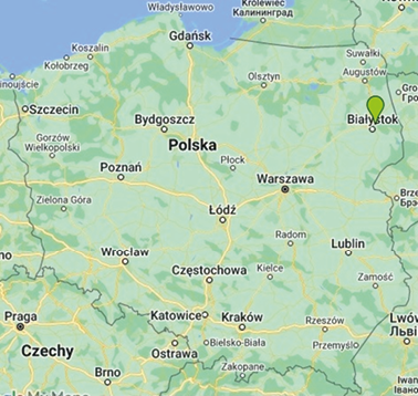
All analysed specimens came from a cemetery located in Dąbrówki (Poland) (Fig. 1). The name „Dąbrówki” appeared for the first time in 1559 (due to the presence of oaks in the local part of Podlasie). The cemetery with skeleton burials was discovered by accident during earthworks in 2018. The cemetery is located on the hill called „Cygańska Brama”. Historical sources do not contain any information about the cemetery (Wawrzeniuk 2021a). The burial equipment found in the cemetery allowed for its preliminary dating pointing to the beginning of the 17th century. It cannot be ruled out that in the remaining areas of the cemetery there are burials from both younger and older periods (Wawrzeniuk 2021b).
Fig. 1. The location of the Dąbrówki village (green drop)
Research work has revealed 62 graves most of which contained adults. The few children’s burials, who were usually buried with adults (Wawrzeniuk 2021a, 2021b). The samples used in this study are part of the osteological collection belonging to the Institute of Biological Sciences Cardinal Stefan Wyszynski University (Poland).
Standard anthropological methods were applied to determine the age and sex of the individuals. The sex of the individuals was determined using methods previously applied by Phenice (1969) and Buikstra, Ubelaker (1994). This includes visual assessments of pelvic and cranial features. The age-at-death of the individual was evaluated based on changes in the morphology of the pubic symphysis using the Todd’s method for changes in pubis symphysis, Brooks and Suchey (1990) standards for changes in the topography of the auricular surface (Buikstra, Ubelaker 1994).
The skeletons of 160 individuals were examined (54 from the 2019 season, 75 from the 2021 season and 31 from the 2022 season). Only 50 of the 160 individuals had complete, undamaged sacral bones, and only these individuals were included in the final study. Table 1 summarises the number of skeletal material from Dąbrówki analyzed in the present study.
| N | |
| Males | 26 |
| Females | 19 |
| Adults* | 3 |
| Subadults | 2 |
| All group | 50 |
N – number of tested individuals; * – unspecified sex; only the division into adults and children was considered, without division into exact age classes).
SBO was analyzed in the study material according to the methods proposed by Buikstra, Ubelaker (1994: 122). Three types of changes were noted: (0) no cleft – all vertebral arches fused, complete; (1) partial cleft – in at least one vertebrae vertebral arch did not fuse with incomplete development or their complete absence; (2) complete cleft – in all sacral vertebrae vertebral arches not fused (incomplete development of vertebral arches or the lack of the vertebral arches) (Fig.2). A word cleft contains any deformity of vertebral arches from discontinuity to the completely lack of these anatomical structures.
In paleoanthropological studies we used differing classification systems for SBO assessment (see Discussion) what may account for variability in reported frequencies. Therefore, the present study was based on the medical assumption stating that SBO at the level of the vertebrae S3, S4, S5 is treated as morphological variability within normal limits (Kumar, Tubbs 2011); the cleft observed at the height of the above-mentioned vertebrae was treated here as no cleft (see Abera et al. 2021; Kim et al. 2018). This allows for the development of methodological standards for historical populations analyses, which not only are necessary for making inter-populations comparisons, but also allow for the comparison of the obtained results to medical data.

Fig. 2. Normal sacral bone (a), sacrum with partial (b) and complete (c) spina bifida oculta (photos by Wolski Adrian)
Statistical significance of the differences in the frequency of the SBO between males and females in the analysed skeletal material was determined using the unilateral test for two components of the structure (a test based on a number of cases and frequencies data). Differences were considered to be statistically significant when p<0.05. All analyses were performed using the Statistica 13.3 software.
The bones of 50 individuals from the skeletal cemetery in Dąbrówka were analysed. SBO was recorded in nine individuals, which consists of 18% of the studied population. Only one of them had a complete cleft of the sacrum (2%). In the remaining cases, we were dealing with incomplete SBO (16%).
In females, no case of complete cleft, and one case of partial cleft was recorded (5%). In males, SBO was found in 7 out of 26 examined skeletons (27%), with a complete cleft in one individual (4%), and a partial cleft in 6 individuals (23%). No cases of SBO (0%) were detected in adults whose sex could not be determined. One case of cleft, a partial cleft (50%), was detected in a subadult. Data on the frequency of SBO in the population from Dąbrówki are presented in Table 2.
| N/n (% n) | |||
| Comlete | Partial | All | |
| Males | 26/1 (4%) | 26/6 (23%) | 26/7 (27%) |
| Females | 19/0 (0%) | 19/1 (5%) | 19/1 (5%) |
| Adults* | 3/0 (0%) | 3/0 (0%) | 3/0 (0%) |
| Subadult | 2/0 (0%) | 2/1 (50%) | 2/1 (50%) |
| All group | 50/1 (2%) | 50/8 (16%) | 50/9 (18%) |
N – numer of individuals; n – numer of individuals with spina bifida oculta; * – unspecified sex.
The next step of the analysis was to compare the significance of differences in the frequency of SBO in males and females. As the results presented in Table 3 show, males (27%) from Dąbrówki show a significantly higher frequency of SBO compared to females (5%).
| Sex | N/n (% n) | p |
| Males | 26/7 (27%) | 0,028 |
| Females | 19/1 (5%) | |
N – numer of individuals; n – numer of individuals with spina bifida oculta; p≤0,05.
In the population from Dąbrówki, spina bifida oculta was recorded in 18% of individuals. Comparing the SBO data in the material from Dąbrówki with other available data from similar historical periods and different regions of Europe, it turns out that the frequency of SBO in the studied material takes intermediate values ranging from 2% (Simalcsik et al. 2011) to 60% (Pliszka 2018).
Although the statistical significance of differences was not assessed here, the observed interpopulation differences and similarities in SBO prevalence may be caused by a number of factors, such as the lack of uniform, standard methods for SBO assessing. For instance, some studies has treated cleft on the height S3-S5 as SBO as a norm and did not take it into consideration (Kim et al. 2018). Similarly, some studies did not provide an information about S3-S5 region treating (e.g., Mays 2006; Silva-Pinto et al. 2010).
| Population | Chronology | % (n/N) | Reference |
| Dąbrówki | XVI | 18% (9/50) | Own data |
| Radom (Poland) | XVII-XIX | 60% (44/74) | Pliszka (2018) |
| Devín-Hrad (Słowacja) | XI-XII | 24% (26/109) | Masnicova, Benus (2003) |
| London | XVIII-XIX | 15% | Saluja (1988) |
| Jassy (Romania) | XVI-XVIII | 2% (2/129) | Simalcsik (2011) |
| Brittany (France) | High Middle Ages | 10% (3/30) | Zemirline et al. (2013) |
N – numer of individuals; n – numer of individuals with spina bifida occulta.
Researchers used to take different segments of the spine into analyses (Hussien et al. 2009; Saluja 1988; Zemirline et al. 2013). There are also studies that did not report any information regarding SBO assessment methods (e.g., Estebaranz-Sánchez et al. 2018). All of this might have contributed to an over- and/or underestimation of the results, and inability to make reliable comparisons between populations. Without clear, unified methods of SBO assessment, it is very difficult to correctly assess and interpret both similarities and differences in SBO prevalence.
Skeletal groups come from different chronological periods as well as different regions of the world. Therefore, their structure might have been influenced by different environmental stress factors and exhibit different genetic endowment adapted to different environmental (socio-economic and biological) conditions (Albrecht et al. 2006). It could be a reason of the observed differences in SBO frequencies. Several studies have found evidence that SBO is associated with a maternal folate (vitamin B9) and folic acid deficiency during the embryonic period (Armstrong et al. 2013; Au et al. 2010; Bentley et al. 2006; Manso, Matos 2023; Melintenda et al. 2023; Pepe et al. 1999). Although the subject of diet in past human populations is still opened, a recent study by Mutlu and colleagues (2020) showed that a Mediterranean Byzantine population, which had access to folate-rich food products, had a lower prevalence of SBO than compared to Anatolian populations. Therefore, it could be assumed that the differences in diet could have influenced the observed interpopulation differences/similarities in SBO prevalence. Proving this hypothesis requires further in-depth research on this research area. Kely and Henneberg (2022) showed an increase of SBO frequency after 1980 despite the introduction of folate supplementation, indicating microevolutionary increase and secular trend in SBO development. This suggests that SBO potentially does not follow the same etiological and embryonic trajectory as spina bifida cistica and has a separate cause. Solving this problem requires further research.
In the population from Dąbrówki SBO was reported more often in males than in females (5%) (Table 3). Similar results were obtained in populations from Iasi (Romania) (5% males, 2% females) (Simalcsik et al. 2011), Devín-Hrad (Slovakia) (males 26%, females 19%) (Masnicova, Benus 2003). In compared populations SBO was more frequently observed in males, and in some populations the condition was not recorded in female group (Shabana et al. 2014; Groza et al. 2013; Saluja 1988; Simalcsik et al. 2011; Masnicova, Benus 2003; Henneberg, Henneberg 1999; Lee 2022; Sarry, Banna 2006). Most studies do not present data on the significance of the difference in the occurrence of SBO between sexes, which makes it difficult to draw final conclusions about the occurrence of the differences. According to the clinical literature, SBO is less common in men than in women, although, some studies show opposite results, indicating SBO more common in men (Eubanks, Cheruvu 2009; Vannier et al. 1981). The reason of such results is not clear yet.
Many factors may have influenced the results obtained in this study as well as other studies (e.g., regarding the higher incidence in males), such as the specificity of the skeletal material (incompleteness, bone damage, small sample size). There are also difficulties in sex determination in skeletal material. Damaged basic diagnostic bony traits, incomplete skeletal material which is often insufficient knowledge about the biology of an examined skeletal group and an influence of environmental stress on skeletal traits used in sex determination make sex determination not always reliable. Moreover, there are some medical studies showing a male excess in cases with lower spinal lesions, and a female excess in upper spinal lesions (Mariman et al. 1992; Seller 1987). These studies, as well as damage or absence of vertebrae of the upper and middle spine, and therefore the analyses of SBO within the sacrum only, may result in a higher frequency of the disorder in males in the studies of past skeletal populations. Determining the reason for these discrepancies raises the need for further research.
Although cases of sacral SBO from the archaeological record have been still reported, there is a lack of consensus in interpretation of spina bifida in skeletal materials (Kumar, Tubbs 2011). One of the reasons is the lack of a standardised methodology of SBO assessment in skeletal remains (Kumar, Tubbs 2011), what causes the under- or overestimation of the results. Another reason is insufficient knowledge about the etiology of the disorder, and therefore an influence of the genetic or/and environmental factors on its manifestation. The results derived from past skeletal populations could not be easily related to the results of clinical records for modern populations. SBO could be documented in higher numbers than those suggested by the medical record because of usually minor symptoms the condition is often clinically undetected (Avrahami et al. 1994; Kumar, Tubbs 2011). Given all the above, research on SBO in past skeletal populations should be continued and developed.
The frequency of spina bifida oculta in the population from Dąbrówka was found in nine individuals, which constituted 18% of the studied population. Only one of studied individuals had a complete cleft in the sacrum (2%). In males SBO was reported in 7 of the 26 studied skeletons (27%). Complete cleft was observed in one individual (4%), partial cleft in 6 individuals, of which 23% belonged to males. No case of complete cleft was detected in females (0%), and one case of partial cleft was detected (5%). We found that differences between males and females in the frequency of this skeletal change were statistically significant.
The population from Dąbrówki did not differ in terms of the frequency of SBO from other populations from different historical periods and areas from other parts of the world. Due to a number of limitations, this study does not allow for drawing far-reaching conclusions.
Due to the lack of uniform research methods for SBO assessment, the inability to make interpopulation comparisons and the relatively high prevalence of SBO phenomenon in both ancient and modern populations, and the unclear etiology of this disease, research on SBO should be continued.
Conflict of interest
Authors declared no conflict of interests.
Authors’ contributions
WA – carrying out skeletal analyses, preparation and description of the statistical analyses; MA – planning and supervision of the research, setting a goal, substantive supervision, corresponding author; WJ – preparation and description of the archaeological part; TJ – content supervision.
Abera Z, Girma A, Bekele A, Oumer M. 2021. Assessment of morphological and morphometrical variations of sacral hiatus in dry human sacra in Ethiopia. Local Reg Anesth 14:25–32. https://doi.org/10.2147/LRA.S277556
Albrecht TL, Scutter SD, Henneberg M. 2006. Radiographic Method to Assess the Prevalence of Sacral Spina Bifida Occulta. Clinical Anatomy 19:000–000. https://doi.org/10.1002/ca.20367
Armstrong S, Cloutier L, Arredondo C, Roksandic M, Matheson C. 2013. Spina bifda in a pre-Columbian Cuban population: A paleoepidemiological study of genetic and dietary risk factors. Int J Paleopathol 3:19–29. https://doi.org/10.1016/j.ijpp.2013.01.004
Au KS, Ashley-Koch A, Northrup H. 2010. Epidemiologic and genetic aspects of spina bifda and other neural tube defects. Dev Disabil Res Rev 16(1):6–15. https://doi.org/10.1002/ddrr.93
Avrahami E, Frishman E, Fridman Z, Azor M. 1994. Spina bifida occulta of S1 is not an innocent finding. Spine 19(1):12–15. https://doi.org/10.1097/00007632-199401000-00003
Banta JV, Lin R, Peterson M, Dagenais T. 1990. The team approach in the care of the child with myelomeningocele. J Prosthet Orthot 2(4):263–273.
Barnes E. 1994. Developmental Defects of the Axial Skeleton in Paleopathology. University Press of Colorado.
Bentley TG, Willett WC, Weinstein MC, Kuntz KM. 2006. Population-level changes in folate intake by age, gender, and race/ethnicity after folic acid fortification. Am J Public Health 96(11):2040–2047. https://doi.org/10.2105/AJPH.2005.067371
Brooks ST, Suchey JM. 1990. Skeletal age determination based on the os pubis: a comparison of the Acsádi-Nemeskéri and Suchey-Brooks methods. Hum Evol 5:227–238. https://doi.org/10.1007/BF02437238
Buikstra J, Ubelaker DH. 1994. Standards for data collection from human skeletal remains. Fayetteville: Arkansas Archaeological Survey, No. 44: Fayetteville.
Copp AJ, Greene ND. 2012. Neural tube defects – disorders of neurulation and related embryonic processes. Wiley Interdisciplinary Reviews: Developmental Biology 2(2):213–227. https://doi.org/10.1002/wdev.71
Estebaranz-Sánchez F, Martínez LM, Alrousan M, Chamel B, Molist M, Coqueugniot E, Pérez-Pérez A. 2018. Spinal dysraphism at the Syrian Neolithic site of Dja’de el-Mughara. Archaeol Anthropol Sci. 10:1375–1387. https://doi.org/10.1007/s12520-016-0460-7
Eubanks JD, Cheruvu VK. 2009. Prevalence of sacral spina bifida occulta and its relationship to age, sex, race, and the sacral table angle. Spine 34:1539–43. https://doi.org/10.1097/BRS.0b013e3181a98560
Fidas A, MacDonald HL, Elton RA, McInnes A, Brown A, Chrisholm GD. 1988. Neurophysiological measurements in patients with genuine stress incontinence of urine and the relation of neurogenic defects to the presence of spina bifida occulta. Brit J Urol 62(1):46–50. https://doi.org/10.1111/j.1464-410x.1988.tb04264.x
Fornoff JE, Egler T, Shen T. 2004. Prevalence of Neural Tube Defects in Illinois 1989-2002. In Epidemiological Report Series, 04:02, Springfield, IL: Illinois Department of Public Health.
Groza VM, Simalcsik A, Bejenaru L. 2013. Spina bifida occulta in medieval and post-medieval skeletons from Iasi City, in North-East Romania. Biologie animala 101–113.
Henneberg RJ, Henneberg M. 1999. Variation in the closure of the sacral canal in the skeletal sample from Pompeii, Italy, 79AD. Perspect Hum Biol 4:177–188.
Holmes LB, Driscoll SG, Atkins L. 1976. Etiologic heterogeneity of neural-tube defects. N Engl J Med 294(7):365–369.
Hussien FH, Sarry El-Din AM, El Samie Kandeel WA, El Banna RAES. 2009. Spinal Pathological Findings in Ancient Egyptians of the Greco-Roman Period Living in Bahriyah Oasis. Int. J. Osteoarchaeol. 19:613–627. https://doi.org/10.1002/oa.984
Josan V, Morokoff A, Maixner WJ. 2008. Epidemiology and aetiological factors. In Memet, Ö.M., Cinalli, G., Maixner, W.J. (coord.), The Spina Bifida. Management and Outcome, Springer-Verlag Press, 59–65.
Kelty ER, Henneberg M. 2022. Sacral spina bifida occulta: A frequency analysis of secular change. Anthrop Rev 85(2):13–62. https://doi.org/10.18778/1898-6773.85.2.02
Kim I, Hopson B, Aban I, Rizk EB, Dias MS, Bowman R, Ackerman LL, Partington MD, Castillo H, Castillo J, Peterson PR, Blount JP, Rocque BG. 2018. Treated hydrocephalus in individuals with myelomeningocele in the National Spina Bifida Patient Registry. J Neurosurg Pediatr 22(6):646–651. https://doi.org/10.3171/2018.5.PEDS18161
Kim IS, Kim H, Hong JH, Lee HJ, Kim MJ, Shin DH. 2018. Lumbosacral Defects in a 16th–18th Century Joseon Dynasty Skeletal Series from Korea. Hindawi BioMed Research International 2018:1–7. https://doi.org/10.1155/2018/7406797
Korsvik HE, Keller MS. 1992. Sonography of occult dysraphism in neonates and infants with MR imaging correlation. Radiographics 12(2):297–306. https://doi.org/10.1148/radiographics.12.2.1561418
Kumar A, Tubbs RS. 2011. “Spina bifida: A diagnostic dilemma in paleopathology.” Clin Anat 24(1):19–33. 24(1):19–33. https://doi.org/10.1002/ca.21058
Lee S. 2022. “An Osteological Study of Spina Bifida in the Nariokotome Homo erectus Skeleton”. Anthropology Senior Theses. Paper 219.
Lee YC, Solomon LB, Ruhli FJ, Schiess R, Ohrstrom L, Sullivan T, Alkadhi H, Henneberg M. 2011. Confirmation of microevolutionary increase in spina bifida occulta among Swiss birth cohorts. Eur Spine 20:776–80. https://doi.org/10.1007/s00586-010-1519-2
Louie K, Irwin B, Thiessen P. 2008. A Booklet on Spina Bifida, An agency of the Provincial Health Services Authority, BC Children’s Hospital, Vancouver.
Manso MT, Matos VMJ. 2023. Spina bifda, the normal, the pathological and the inbetween: frst evidence from a forensic osteological collection. Int J Legal Med. https://doi.org/10.1007/s00414-023-03066-2
Mariman EC, Hamel BC. 1992. Sex ratios of affected and transmitting members of multiple case families with neural tube defects. J Med Genet 29:695–698. https://doi.org/10.1136/jmg.29.10.695
Masnicova S, Benus R. 2003. Developmental Anomalies in Skeletal Remainsfromthe Great Moravia and Middle Ages Cemeteries at Devín (Slovakia). Int J Osteoarchaeol 13(5):266–274. https://doi.org/10.1002/oa.684
Mays S. 2006. Spondylolysis, Spondylolisthesis, and Lumbo-Sacral Morphology in a Medieval English Skeletal Population. Am J Phys Anthropol 131:352–362. https://doi.org/10.1002/ajpa.20447
McComb JG. 1997. Spinal and cranial neural tube defects. Seminars in Pediatric Neurology 4(3):156–166. https://doi.org/10.1016/S1071-9091(97)80034-4
Melintenda S, Varotto E, Pappalardo E, Guzzardi L, Papa V, Palermo D, Galassi FM. 2023. Spina Bifida Sacralis Occulta from Ancient Greek Sicily (Pozzanghera Necropolis, Leontinoi, 6th–4th Century BC): Anatomical, Anthropological and Ethnomedical Considerations on the Insular Presentation of this Congenital Anomaly. Anthropol Rev 86(2):13–25. https://doi.org/10.1016/j.jchb.2010.07.001
Mohd-Zin SW, Marwan AI, Abou Chaar MK, Ahmad-Annuar A, Abdul-Aziz NM. 2017. Spina bifida: Pathogenesis, mechanisms, and genes in mice and humans. Scientifica 1–29. https://doi.org/10.1155/2017/5364827
Moore KL, Persaud TVN. 2018. The Developing Human: Clinically Oriented Embryology. Elsevier.
Mutlu H, Kizgut B, Sözer ÇS, Ürker K, Açar O, Erol AS. 2020. Sacral spina bifida occulta rare occurrence in Byzantine Belen-tepe population in Muğla, Turkey: A possible case for adequate folic acid intake. Homo 71(3):175–188. https://doi.org/10.1127/homo/2020/1233
Northrup H, Volcik KA. 2000. Spina bifida and other neural tube defects. Curr Probl Pediatr Adolesc Health Care 30(10):317–332. https://doi.org/10.1067/mpp.2000.112052
Pepe F, Pepe P, Grillo S, Insolia G. 1999. As-sunzione periconcezionale di acido folico in coppie siciliane a rischio di ricorrenza di DNT. Minerva Ginecol 51(10):399–402.
Phenice WT. 1969. A newly developed visual method of sexing the os pubis. Am J Biol Anthropol 30(2):297–301. https://doi.org/10.1002/ajpa.1330300214
Pliszka AM. 2018. Rozczep kości krzyżowej (spina bifida occulta) a asymetria fluktuacyjna obserwowana na populacji z Radomia (XVIII–XIX wiek) w świetle badań archeologicznych. Master thesis. Unpublished.
Sairyo K, Goel VK, Vadapalli S, Vishnubhotla SL, Biyani A, Ebraheim N, Terai T, Sakai T. 2006. Biomechanical comparison of lumbar spines with or without spina bifida occulta. A finite element analysis. Spinal Cord 44:440–444. https://doi.org/10.1038/sj.sc.3101867
Saluja PG. 1986. Evidence of spina bifida in skeletal remains from Ireland. Ir Med J 79:145–149.
Saluja PG. 1988. The incidence of spina bifida occulta in a historic and a modern London population. J Anat 158:91–99.
Seller MJ. 1987. Neural tube defects and sex ratios. Am J Med Genet 26:699–707. https://doi.org/10.1002/ajmg.1320260325
Shabana A, Asad AA, MBBS, FCPS (Medicine), Saffia S. 2014. The prevalence of spina bifida occulta in Pakistani population: A study of dry human sacra. Anaesth Pain Intensive Care 18(2):157–161.
Silva-Pinto E, Arriaza B, Standen V. 2010. Evaluación de la frecuencia de espina bífida oculta y su posible relación con el arsénico ambiental en una muestra prehispánica de la Quebrada de Camarones, norte de Chile. ev Med Chile; 138:461–469. https://doi.org/10.4067/S0034-98872010000400010
Simalcsik A, Miu G, Groza VM, Simalcsik RD. 2011. Regarding occult spinal dysraphism (spina bifida occulta), focussing especially on a medieval population from Isai. Biol Anim 62:131–141.
Solomon LB, Ruhli FJ, Lee YC, Henneberg M. 2009. Secular trend in the opening of the sacral canal. An Australian study. Spine 34:244–248. https://doi.org/10.1097/BRS.0b013e3181908ca2
Spacca B, Buxton N. 2008. Spina bifida occulta and monozygotic twins. Journal of Neurosurg Pediatr 2(4):258–260. https://doi.org/10.3171/PED.2008.2.10.258
Vannier JP, Lefort J, Cavelier B, Ledosseur P, Assailly C, Feingold J. 1981. Spina bifida cystica families X-Ray examination and HLA typing. Pediatr Res 15:326–329.
Wawrzeniuk J. 2021a. Sprawozdanie wstępne z archeologicznych badań ratowniczych cmentarzyska szkieletowego w miejscowości Dąbrówki, st. 11, gm. Wasilków, woj. podlaskie, AZP 35-87/25 – nazwa lokalna „Cygańska Brama”. Warszawa.
Wawrzeniuk J. 2021b. Zapomniane wiejskie nowożytne cmentarze Podlasia -stan badań a perspektywy badawcze. Saeculum Christianum 28:133–148.
Zemirline A, Vincent JP, Sid-Ahmed S, Le Nen D, Dubrana F. 2013. Lumbo-sacral malformations and spina bifida occulta in medieval skeletons from Brittany. Eur J Orthop Surg Traumatol 23(2):149–153. https://doi.org/10.1007/s00590-012-0967-2

