 https://orcid.org/0000-0002-0605-665X
Introduction
Nowadays, the assessment of the condition of the dentition is a common aspect of bioarchaeological research (Ioannou and Henneberg 2017; Koruyucu and Erdal 2021). This is due to the fact that the odontological material is well preserved at archaeological sites, even where bone materials are subject to significant fragmentation and destruction.
One of the diseases assessed in bioarchaeological materials is dental caries (Yanko et al. 2017; Moles 2023). Based on the analysis of the frequency of dental caries, eating habits and/or the level of hygiene can be assessed indirectly. However, it must be remembered that oral hygiene and eating habits are only one of the cariogenic factors. Clinical practice indicates that other causative factors may be congenital hypomineralization of enamel, malocclusion, tooth displacement in alveolar arch or the chemical composition of saliva (Fejerskov and Kidd 2003). However, in the historical materials, not all of the indicated factors are possible to determine.
Additionally, the results of odontological research can be interpreted in the context of archaeobotanical, archaeozoological or isotopic research (Tomczyk et al. 2020a; Schata et al. 2022). This information is crucial for understanding the economic and social status of studied populations. Therefore, in the odontological studies of historical populations, caries studies play an important role.
In this work, attention will be focused on the period of the early Middle Ages in Poland. In the period up to 1138, in the lands between the Oder and the Bug Rivers a centralized state ruled by the Piast dynasty was established, which were known as ‘Poland’ from the 10th and 11th centuries (Urbańczyk 2008). Although quite a lot of early medieval burial grounds in Poland have been reported (e.g., Cracow, Wroclaw, Milicz) (Miszkiewicz and Gronkiewicz 1986; Kwiatkowska 2005; Mietlińska 2015), ‚broadband’ odontological examinations have been only carried out in selected ones (e.g., Cedynia, Ląd, Brześć Kujawski, Radom) (Kozubkiewicz et al. 1957; Kozubkiewicz and Trachtenberg 1960; Borysewicz and Otocki 1975; Borysewicz-Lewicka and Otocki 1978). However, a lot of these studies dated back to the 1960s and 1970s. This means that each new site from the early Middle Ages examined in terms of odontology is valuable for understanding this period in the history of Poland.
The aim of this study was to assess changes in the diet and level of hygiene among young children (the age of 1–8 years) and adults who inhabited Starorypin (Poland) in the early medieval period, and compare the studied population with other selected populations from early medieval Poland. This study aims to address the following questions: i) what was the frequency of dental caries in children and adults, ii) where were dental caries most often located, iii) in what age groups among children were caries most common and iv) how does the studied group present itself against groups from other sites of the early Middle Ages?
Materials and Methods
Archaeological context
The Starorypin site is located in North Eastern Poland, approximately 65 km from Toruń (Fig.1). Studies of the settlement complex in Starorypin indicate that it was an important settlement in the early and late Middle Ages and an administrative center of the interfluve of the Vistula, Drwęca and Skrwa. The key to research on the history and culture of the early medieval Dobrzyń land has been the discovery and partial recognition of the cemetery from this period (site 6). The location of the necropolis remained unrecognized until recently and the work was focused on former central places and settlements (Lewandowska 2016, 2022).
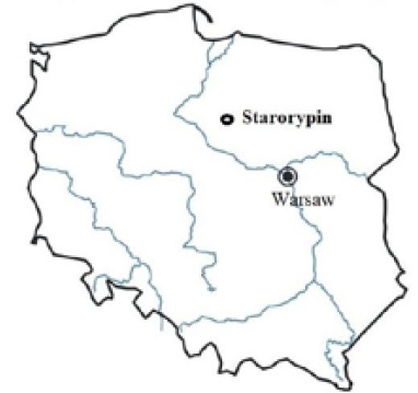
Fig. 1. Location map of the study area
The history of research on early medieval burials in Starorypin dates back to the 1960s. At that time, following heavy rains, human bones used to be observed in the escarpment. As a result of the archaeological intervention, six very poorly preserved skeletons were recorded (Grześkowiak 1963, 1967). Another accidental unveiling of a fragment of the necropolis took place in 2010, when human remains were found on the heap during a linear investment. As a result of excavations (until 2022) and anthropological analysis, 52 graves and 29 objects classified as bone clusters were recorded. Within the excavated graves, 68 individuals were identified, while 29 individuals were identified in the group of loose bones.
The fragment of the necropolis examined has provided, apart from human bones in various states of preservation, a representative historical material, constituting an equipment and personal belongings of the deceased, indicating various fields of production and material manifestations of everyday life and spiritual culture. In particular, numerous grave inventories have been discovered in the burials of adult women. The largest group of all items have been ornaments, primarily necklaces made of glass beads and other materials, including semi-precious stones (carnelian, amethyst, rock crystal) and amber. The findings also included temple rings and other rings. Following ornaments, the second largest group of items have been tools and everyday objects.
The observed funeral rite (orientation of graves, skeletal burial) can be associated with Christian eschatology. On the surface of the necropolis examined to date, the confirmed form of the grave was a flat grave (i.e., a grave devoid of any superstructures visible on the surface). The basic feature of the layout of early medieval skeletal cemeteries is the row arrangement of graves, which is also observed at the Starorypin site (Bojarski 2020; Stawska 2003; Stawska et al. 2010).
Based on the typologies of objects deposited in the graves and the analysis of the obtained radiocarbon dates, fragments of ceramic vessels and reflections on the functioning of the entire settlement complex, including the stronghold and the suburban settlement, it was possible to determine the time of functioning of the cemetery, which was operating from the first half of the 11th century to the second half of that century and into the 12th (Błędowski and Lewandowska 2022).
Dental materials
A total of 25 adults (males: 12, females: 13), with a total of 346 permanent teeth (males: 181, females: 165), and 14 children, with a total of 131 deciduous teeth were examined.
Regarding adults, the basic criterion for material selection was the possibility of establishing the sex and age-at-death of the individuals. The sex and age-at-death of individuals were assessed according to the commonly accepted anthropological methods (Brooks and Suchey 1990; Buikstra and Ubelaker 1994).
The examined children were divided into three age groups: 1.0–2.5 years, 3.0–5.5 years and 6.0–8.0 years. These age groups reflect the successive stages of tooth eruption (Liversidge and Molleson 2004). Because among children no permanent teeth have been preserved, only the deciduous dentition was used.
The diagnosis of dental caries was carried out through visual, radiographic and fluorescent techniques:
(i) Visual observation of the dental caries was conducted using a 3x dental magnifying glass and a sharp dental probe. Visual inspection was performed under a direct dental unit light.
(ii) We used radiographic techniques for diagnosis; for this task, a portable X-ray machine (EZX-60, Edlen Imaging, USA) was used.
(iii) A light-induced fluorescence technique was used for the detection of initial enamel caries on occlusal and approximal surfaces. For this task, we used the VistaCam iX Proof device (Dürr Dental, Germany).
The location of carious lesions (approximal, root and occlusal) was considered and analyzed. When more than one surface was affected, each surface was analyzed separately.
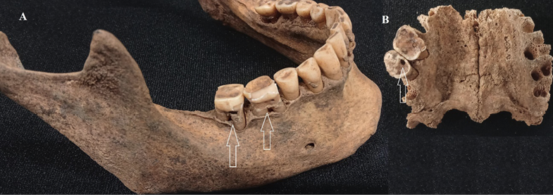
Fig. 2. Examples of the advancement of caries in adults (A: No 61(1)/2022, B: No 61/2022)
In the deciduous dental analyses, particular attention was paid to early childhood caries (ECC). This kind of caries appears in caries-resistant regions, such as the labial surfaces of the upper incisors, the upper and lower molars and, more rarely, in the upper canine and (even more rarely) in the lower canine and incisors (Çolak 2013; Begzati et al. 2015).
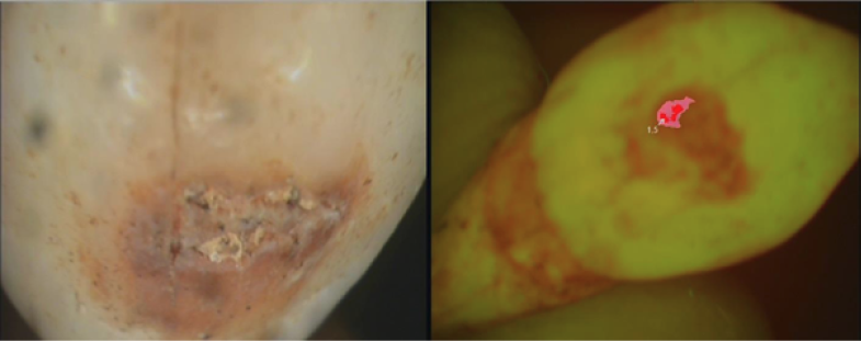
Fig. 3. Caries on the labial surface of the deciduous canine (FDI: 83) macroscopic photo and using fluorescent light (No: 35/2020)
The frequency of caries (i.e., the number of teeth affected by caries) was calculated based on the proportional correction factor (PCF) following Erdal and Duyar (1999, 2003). The PCF was applied because dental materials are often missing in historical materials (e.g., during post-mortem deposition). Ofter, the anterior teeth are missing due to their morphological structure. The PCF provides caries rates of anterior and posterior teeth according to their appropriate numbers: i) permanent teeth: three-eighths for anterior and five-eighths for posterior teeth; ii) deciduous teeth: three-fifths for anterior and two-fifths for posterior teeth, multiplied by the number of caries observed.
The analyses also included an assessment of the severity of carious lesions. Carious lesions were divided into (i) enamel decay (early-stage tooth decay, which is formed before a cavity) and (ii) cavities.
In order to estimate the intra-observation and inter-observation error, 80 teeth were chosen at random. These analyses were performed by two experienced odontologists (JT, DO-K).
The chi-squared test was used to analyze the differences in caries frequency. In addition, due to the small samples, the Fisher’s Test was used. The chi-squared test was also used to compare measurements in the intra- and inter-observation error estimations. Statistical analyses were performed using the R software (http://www.R-project.org. 2013). Differences with p < 0.05 were considered statistically significant.
Results
The intra- and inter-observation error was found to be statistically insignificant (p = 0.6732 and p = 0.6223, respectively).
Out of 25 adults, 20 were diagnosed with dental caries, while caries were observed in 4 of the 14 children. Dental caries were recorded in 33% (115/346) of the permanent teeth and in 10% (13/131) of the deciduous teeth. Dental caries were found in 26% (43/165) of females and 40% (72/181) of males; however, the observed differences are not statistically significant (p = 0.0912).
Dental caries in adults were diagnosed most frequently on the approximal surfaces and least often on the root. In contrast, in children, dental caries were most often diagnosed on the occlusal surface. In both cases, the observed differences were statistically significant.
Analyzing children in successive age categories, a clear increase in dental caries in the second age category (3.0–5.5) was observed. Changes in dental caries frequency between age categories were statistically significant (Fisher Test: p = 0.0056, Monte-Carlo simulations: p = 0.0040). Although there were not many tested individuals, ECC was diagnosed on all deciduous teeth of children aged 3.0–5.5 years (Table 1).
| Total | anterior | posterior | ECC# | approximal | root | occlusal | |
| Adult | 115/346 (33%) |
2/115 (0.65)* |
113/115 (61.4)* |
- | 79/115 (69%) |
27/115 (23%) |
46/115 (40%) |
| p < 0.0001 χ2 = 48.852 | |||||||
| Children | 13/131 (10%) |
4/13 (18.4)* |
9/13 (27.6)* |
13/13 (100%) |
6/13 (46%) |
0/13 (-) |
11/13 (85%) |
| p < 0.0001 χ2 = 18.979 | |||||||
| 1.0-2.5 | 1/58 (2%) |
0/1 (-) |
1/1 (40)* |
1/1 (100%) |
1/1 (100%) |
0/1 (-) |
0/1 (-) |
| 3.0-5.5 | 12/61 (20%) |
2/12 (10)* |
10/12 (33)* |
12/12 (100%) |
6/12 (50%) |
0/12 (-) |
10/12 (83%) |
| p = 0.0002 χ2 = 17.1 | |||||||
| 6.0-8.0 | 0/12 (-) |
0/0 (-) |
0/0 (-) |
0/0 (-) |
0/0 (-) |
0/0 (-) |
0/0 (-) |
*proportional correction factor (PCF).
# early childhood caries (ECC).
| Archaeological site | Period | Frequency of teeth with dental caries | References |
| Kraków-Zakrzówek | 11th–13th c. | 3.1% | Gleń 1975 |
| Brześć Kujawski | 11th c. | 9.8% | Kozubkiewicz et al. 1957 |
| Kałdus | 11th–12th c. | 10.4% | Kozubkiewicz and Trachtenberg 1960 |
| Garbary | 12th–13th c. | 12.0% | Borysewicz-Lewicka and Otocki 1978 |
| Cedynia | 11th–13th c. | 13.1% | Stopa and Perzyna 1978 |
| Ląd | 11th–13th c | 14.0% | Borysewicz and Otocki 1975 |
| Stary Brześć | 12th–16th c. | 14.0% | Borysewicz and Otocki 1975 |
| Ołbin | 12th–13th c. | 14% | Kwiatkowska 2005 |
| Wrocław | 12th–13th c. | 15.7% | Staniowski et al. 2011 |
| Wrocław – pl. Dominikanski | 12th–14th c. | 22% | Kwiatkowska 2005 |
| Radom | 11th–12th c. | 38.0% | Tomczyk et al. 2020b |
| Starorypin | 11th–12th c. | 33.0% | presented research |
In this study, an attention was paid to the degree of advancement of dental caries, separating enamel decay from cavities. On the permanent teeth, 23 (20%) enamel changes and 96 (83%) cavities were observed, while on the deciduous teeth, 8 (62%) cases of enamel decay and 5 (38%) cavities were observed.
Discussion
Compared to data obtained from other archaeological sites from the early Middle Ages in Poland, the occurrence of dental caries in Starorypin is considerably higher (Table 2). This may be due to a small sample size, although it cannot be ruled out that the low frequency of teeth with caries results from a different methodology, e.g. an examination based only on macroscopic assessment, without the use of a fluorescent camera or X-ray. Lucas et al. (2010) showed that the use of the radiological method increases the detection of caries. Also, Tomczyk et al. (2014), showed that the use of micro-CT and VistaCam iX Proof increases the detection of early dental caries. Comparative studies have shown that these methods of dental caries detection were not used, which could to some extent underestimate the detection of the disease, especially in its early stages. This supposition may be supported by the comparability of the results from Starorypin with another Polish early medieval population, such as the Radom population, which was studied using the same devices and a similar methodology (Tomczyk et al. 2020b). In the early medieval population from Radom, for example, dental caries were identified in 38% of permanent teeth.
The assessment of the frequency of dental caries is used in archaeological research to reconstruct living conditions, health and/or hygiene habits of past populations (Yanko et al. 2017; Moles 2023). Although the frequency of teeth infected with caries seems to be significantly high at the Starorypin site, it should be remembered that this level was clearly higher in subsequent historical periods. The high frequency of teeth infected with caries observed at at the Starorypin site has been explained by a monotypic economy and a cariogenic diet (O’Sullivan et al. 1993; Kurek et al. 2009; Bertilsson et al. 2022).
The reconstruction of the diet of the Starorypin population was made on the basis of archaeozoological and archaeobotanical analyses. This research showed a diverse picture of the environment, which was probably resulted in a high diversity of the produced foods. For example, it was shown that the local population not only raised mammals (cattle, pigs, sheep) and poultry but was also engaged in hunting, as evidenced by the presence of deer, roe deer and wild boars and fishing (pike, carp and tench) (Makowiecki 2022). Fertile areas were dominated by deciduous forests with a predominance of hornbeam and poor sandy sites that were covered with pine and pine-oak forests. On the other hand, wetlands around water reservoirs and swamps were occupied by communities dominated by alder. Lands used to be acquired not only for crops and pastures, but also for housing estates and roads (Noryśkiewicz 2022). The conducted analyses show that diet of the early medieval population was not limited to cariogenic products, such as cereals. Instead, this population exhibited a rather varied diet containing a large proportion of animal protein. High-content protein diets have been shown to reduce the acidity of the saliva and, thus, neutralize the decalcifying acids that cariogenic bacteria produce. The consumption of diets that are relatively high in protein increases blood urea levels, which may lead to relatively high salivary urea levels. Since urea appears to be the main substrate for the formation of dental plaque, a slight increase in the salivary urea concentration or output may reduce the development of caries (Dawes 1970; Silverstone et al. 1981; Zabokova-Bilbilova et al. 2012). However, research on diet composition in the above studies requires further isotope studies to confirm this interpretation.
In this study, attention was focused on the location of dental caries. In the studied population, dental caries dominated on the approximal surfaces. This can be explained by intensive chewing, conditioned by the type of food, which potentially led to a strong abrasion of the occlusal surfaces. This deprived the teeth of numerous retention places where food could linger, destroying the crown of the tooth, while the poor level of hygiene meant that the remains of the food were in the cervical regions long enough to undergo fermentation and start the process of enamel destruction. Similar observations apply to other early medieval populations from Poland, such as Cedynia, Słaboszewo and Góra Chełmska (Borysewicz and Otocki 1975; Stopa and Perzyna 1978; Torlińska-Walkowiak and Jerszyńska 2011).
The examination of the degree of dental caries advancement showed that carious lesions were diagnosed in the majority. Given that the age of most individuals at the time of consent was not high (20/35 years old) (Myszka et al. 2022), it can be assumed that dental caries had either an acute course or developed at a very early age. Unfortunately, observations of the severity of caries were carried out sporadically in populations from the early Middle Ages. However, a similar observation was reported on the early medieval population from Chełmska Góra (Borysewicz and Otocki 1975) and Garbary (Borysewicz-Lewicka and Otocki 1978). Interestingly, in these studies enamel decay was most frequently observed among children. It can be assumed that the development of these changes was halted by the death of the individual.
In the analyzed population of Starorypin, children with deciduous dentition were also included. Overall, dental caries were diagnosed in 10% of children. Due to the scarcity of archeological studies on children, it is rather difficult to compare the results of our study to other early medieval sites in Poland. In Garbary (12–13th c.), dental caries were diagnosed on the deciduous teeth of children in the infans I category at the level of 3% (Borysewicz-Lewicka and Otocki 1978). Equally, low dental caries on deciduous teeth were recorded on the primary teeth of children from Chełmska Góra (Borysewicz and Otocki 1975). In the infans I group from Cedynia, the total frequency on the tooth type level of carious lesions was 2.8% (Torlińska-Walkowiak and Jerszyńska 2011). However, the cited studies did not divide the tooth material into three age categories, but, instead, analyzed the tooth material as one age group of infans I. This certainly does not facilitate comparative analyses.
Dental caries in the deciduous dentition allows for inferences on weaning and feeding practices that promote dental decay. For instance, a lower frequency of carious lesions suggests a simpler diet based on low in cariogenic products. The Starorypin population may have had more access to processed and sweetened foodstuffs and, therefore, refined carbohydrates were more readily available. In our study dental caries were most often observed among children aged 3.0–5.5 years. This would suggest that the process of introducing solid products started before the age of three, which corresponds with population isotope studies from the early Middle Ages in Radom showing that the weaning period occurred roughly between 1.1 and 2.5 years of age (Tomczyk et al. 2021). However, the presented interpretation must be confirmed by isotope studies.
The dental caries among children were diagnosed on the occlusal surfaces, which, due to a slight abrasion, exhibited many furrows, providing places for the accumulation of food remains. Similar observation regarding the location of caries on deciduous teeth was reported in the Góra Chełmska site (Borysewicz and Otocki 1975).
When examining the teeth of non-adults, it is worth paying attention to early childhood caries (ECC) (Çolak 2013; Begzati et al. 2015). This kind of dental caries is also known as „severe early childhood caries” or „nursing caries”. Newly erupted teeth do not have fully matured enamel; it is thinner, with large dental tubules. In addition, compared to adults, salivation is slower in children (Rohnbogner and Lewis 2016) and decay processes develop quicklier. Therefore, the development of ECC is characterized by highly dynamic changes, which, in a short time, lead to the destruction of the tooth crown and, consequently, to the development of diseases of pulp and periapical tissues. It is generally accepted that ECC develops by the age of 5 years. The dynamics of changes in ECC are impossible to detect in bioarcheological materials because it is not possible to determine the rate of decay. However, their location can be helpful for identification. This kind of caries appears in caries-resistant regions: labial surfaces of the upper incisors, in the upper and lower molars and, more rarely, in the upper canine and (even more rarely) in the lower canine and incisors (Çolak 2013; Begzati et al. 2015). Although the material in our study is sparse, ECC can be observed primarily among individuals aged 3.0–5.5, although ECC was also observed in the earlier period (1.0–2.5 years).
Conclusion
Our examination of the masticatory apparatus, narrowed down to the assessment of dental caries, is an important element of bioarchaeological research. The paper presents analyses performed on the early medieval population discovered in Starorypin. Although the discovered burials are not numerous and require further isotope analyses, they broaden our knowledge about the early medieval population of Poland. Furthermore, the study investigated not only the permanent dentition of adults, but also the deciduous dentition of children.
The presented analyses show that the population of Starorypin probably functioned on a varied diet, which did not generate pro-inflammatory processes. The location of dental caries on the approximal surfaces in adults and on the occlusal surfaces in children corresponds to the pattern which was also observed in other populations from this period.
Conflict of interest
The Authors do not have any conflict of interest.
Authors’ contribution
AM: assessment of the sex and age of the individuals; DO-K: dental analysis, preparation and description of the manuscript; MZ: statistical analysis; WN: statistical analysis; JL: archaeological and historical information; JT: dental analysis, planning and supervision of the research, setting a goal, substantive supervision.

