 https://orcid.org/0000-0002-1780-8992
Introduction
Anthropometry often uses a collection of human bodily measurements in order to understand human physical variation as it plays an important role in plastic surgery, prosthetics, personal identification, biometrics and forensic profiling (Purkait and Singh 2007; Ekanem et al. 2010; Deopa et al. 2013). The ear, or pinna, is an important but under-studied feature of the face which shape and size conveys information about age and sex (Brucker et al. 2003). The shape, size and orientation of each pinna are as unique as fingerprints although it is possible to make some generalizations (Alexander et al. 2011; Osunwoke et al. 2018).
Currently, anthropometric studies have shown that the morphological variation of the external ear depends on age, sex and race and side (i.e., side-to-side variation) (Meijerman et al. 2007; Purkait and Singh 2007; Murgod et al. 2013). Jung and Jung (2003) surveyed the dimensions and characteristics of Korean ears and found that age, sex, and specific ethnic group were contributing factors of auricular dimensions. Sharma et al (2007) carried out a morphometric study in India where it was observed that North-West Indians have smaller ear lobules compared to Caucasian and Japanese populations but similar to those observed among the Onge tribe of Andhra (India) and Newars of Nepal. A study done by Murgod et al (2013) that assessed ear shape and earlobe metric dimensions of 300 Indian adult subjects concluded that identifying males was about 69% accurate while that of females was 72%. Several Nigerian studies have been conducted on the morphological and morphometric variation of an external ear (Ekanem et al. 2010; Eboh 2013; Taura et al. 2013). The aim of this study was to examine the accuracy of prediction of age and sex of a selected Nigerian Igbo population using auricular morphometrics for forensic applications.
Materials and Methods
A cross-sectional, descriptive study design was applied to randomly select Igbo heterogeneous indigenes residing in the South-Eastern states of Nigeria (Abia, Anambra, Ebonyi, Enugu and Imo). Using the Cochran formula, the calculated sample size was 300 adult subjects, comprised of 142 males and 158 females of at least 16 years of age. Study participants showed no past records of ear abnormalities or surgical operations and were selected in line with the Declaration of Helsinki research ethics protocol for human research. Both primary and secondary data were obtained from study participants. Biological profile, such as age and sex, made up the primary data while ear measurements the secondary data.
The participants were made to sit in a Frankfort horizontal position and with the aid of a digital vernier caliper (validated by calibrating to 0.01mm), the ear measurements obtained from participants included the following:
- total ear height (TEH) – The length measured from the most superior part of the ear to the most inferior part of the earlobe.
- ear width (EW) – The length measured from the most anterior to the most posterior parts of the ear.
- lobule height (LH) – The distance between the most inferior point of the earlobe, and the deepest point of the intertragic notch
- lobule width (LW) – The distance between the most anterior and most posterior points of the earlobe
To test the reliability of the instrument, the above measurements were obtained twice and the average score was calculated.
In addition to these parameters, two indices defining the proportion of the ear were also calculated: the ear index (EI) [ear width / ear height x 100] and the lobule index (LI) [lobule height / lobule width x 100].
Statistical Analyses: Raw data obtained from participants were recorded into Microsoft Excel 2019 version and analyzed using the Statistical Package for the Social Sciences (SPSS version 23.0). Sexual dimorphism was determined using the Independent Sample t-test, while side (left and right) differences were determined using the Paired sample t-test. Differences in auricular dimensions between age categories were determined using Analysis of Variance (ANOVA). Confidence interval was set at 95% and therefore P<0.05 was considered significant.
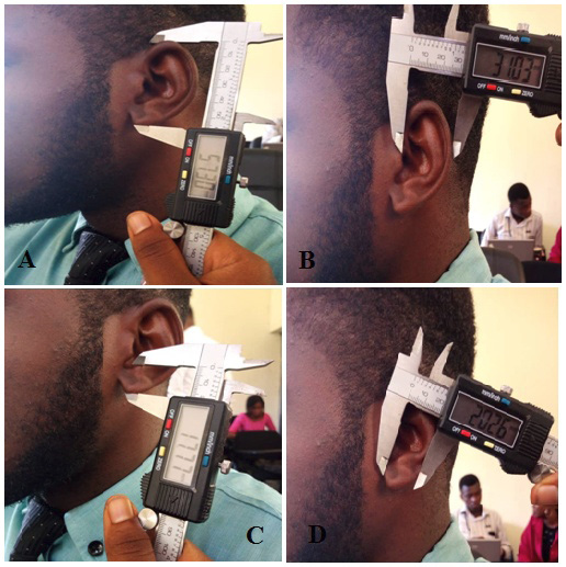
Fig. 1. Ear morphometric dimensions. (A) Total ear height (B) Ear width (C) Lobule height (D) Lobule width
Results
Males had a mean age of 35.34, SD=12.04, while for female subjects, the mean age was 35.21, SD=12.90 (Table 1). The minimum age for males was 16, while the maximum was 62, while for females; it was 16 and 63 respectively (Table 1). There was no significant difference between the mean age of male and female subjects (Table 1).
Gender differences between auricular dimensions are shown in Table 2. Significant differences were observed in left ear height, left lobular height, left lobular width and left ear index (Table 2).
Significant differences in symmetry (i.e., side differences) in male Igbo auricular dimensions were only observed in total ear height (Table 3).
Significant differences regarding asymmetry in female Igbo auricular dimensions were observed in ear width, lobular height, lobular width and ear index (Table 4).
| Sex | Age in years | T-test | ||||||
| N | Min | Max | Mean | S.D | t-value | Df | p-value | |
| Male | 142 | 16 | 62 | 35.44 | 12.04 | 0.09 | 298 | >0.05 |
| Female | 158 | 16 | 63 | 35.32 | 12.90 | |||
| Total | 300 | 16 | 63 | 35.38 | 12.48 | |||
Min = Minimum, Max = Maximum, S.D = Standard Deviation, df = degree of freedom
| Auricular Dimensions (mm) | Male [142] | Female [158] | ||||
| Mean (SD) | Min | Max | Mean (SD) | Min | Max | |
| Right Auricle | ||||||
| Total Ear Height | 47.01 (6.30) | 24.06 | 60.84 | 48.25 (6.83) | 24.06 | 65.14 |
| Ear Width | 25.06 (4.14) | 11.16 | 33.22 | 25.19 (3.93) | 13.45 | 33.51 |
| Lobular Height | 13.92 (2.61) | 10.03 | 20.54 | 14.44 (2.67) | 10.19 | 21.57 |
| Lobular Width | 12.99 (2.30) | 10.00 | 22.15 | 13.37 (2.26) | 10.00 | 20.25 |
| Ear Index | 53.47 (7.23) | 30.97 | 70.45 | 52.50 (6.85) | 30.96 | 69.39 |
| Lobular Index | 95.17 (18.12) | 55.89 | 186.26 | 94.60 (19.59) | 53.48 | 193.44 |
| Left Auricle | ||||||
| Total Ear Height | 46.24 (5.85) | 25.67 | 58.29 | 48.25 (6.24) | 25.67 | 61.66 |
| Ear Width | 24.68 (4.32) | 10.21 | 33.38 | 24.57 (4.21) | 10.12 | 34.05 |
| Lobular Height | 13.99 (2.18) | 10.21 | 21.24 | 14.89 (2.55) | 10.19 | 21.24 |
| Lobular Width | 13.30 (1.94) | 10.11 | 19.75 | 13.78 (2.04) | 10.21 | 19.26 |
| Ear Index | 53.28 (6.69) | 32.48 | 75.12 | 50.93 (7.05) | 29.20 | 72.06 |
| Lobular Index | 96.80 (18.56) | 61.29 | 193.44 | 94.30 (16.63) | 61.80 | 153.45 |
SD = Standard Deviation, M.D = Mean Difference, S.E.M = Standard Error of Mean
|
Auricular Dimensions (mm) |
M.D | S.E.M |
95% C.I of the Difference |
t-value | df | p-value | |
| Lower | Upper | ||||||
| Right TEH - Left TEH | 0.76 | 0.27 | 0.23 | 1.30 | 2.84 | 141 | <0.05 |
| Right EW - Left EW | 0.38 | 0.21 | -0.04 | 0.80 | 1.77 | 141 | >0.05 |
| Right LH - Left LH | -0.07 | 0.22 | -0.51 | 0.36 | -0.34 | 141 | >0.05 |
| Right LW - Left LW | -0.31 | 0.18 | -0.67 | 0.06 | -1.66 | 141 | >0.05 |
| Right EI - Left EI | 0.19 | 0.57 | -0.93 | 1.30 | 0.33 | 141 | >0.05 |
| Right LI - Left LI | -1.63 | 1.96 | -5.50 | 2.24 | -0.83 | 141 | >0.05 |
| Auricular Dimensions (mm) | M.D | S.E.M |
95% C.I of the Difference |
t-value | df | p-value | |
| Lower | Upper | ||||||
| Right TEH - Left TEH | 0.00 | 0.27 | -0.53 | 0.53 | -0.01 | 157 | >0.05 |
| Right EW - Left EW | 0.62 | 0.21 | 0.20 | 1.05 | 2.91 | 157 | <0.001 |
| Right LH - Left LH | -0.44 | 0.19 | -0.82 | -0.07 | -2.32 | 157 | <0.05 |
| Right LW - Left LW | -0.40 | 0.18 | -0.75 | -0.06 | -2.30 | 157 | <0.05 |
| Right EI - Left EI | 1.57 | 0.57 | 0.45 | 2.70 | 2.76 | 157 | <0.05 |
| Right LI - Left LI | 0.30 | 1.86 | -3.38 | 3.98 | 0.16 | 157 | >0.05 |
Age related differences were observed in right ear index, left total ear height, and left ear width as well as left ear index (Table 5).
| Auricular Dimensions (mm) | Age (years) | Descriptive Statistics | ANOVA | ||||||
| N | Min | Max | Mean | S.D | df | F-value | p-value | ||
| Right Auricle | |||||||||
| Total Ear Height | 16 - 25 | 90 | 32.15 | 59.95 | 48.28 | 5.73 | 4 | 2.17 | >0.05 |
| 26 - 35 | 61 | 24.06 | 65.14 | 46.27 | 7.87 | ||||
| 36 - 45 | 78 | 24.06 | 62.84 | 46.86 | 6.36 | ||||
| 46 - 55 | 52 | 32.68 | 61.81 | 49.47 | 6.68 | ||||
| 56 and above | 19 | 39.48 | 60.84 | 47.53 | 5.98 | ||||
| Ear Width | 16 - 25 | 90 | 11.16 | 33.22 | 25.15 | 3.93 | 4 | 1.30 | >0.05 |
| 26 - 35 | 61 | 13.45 | 32.81 | 24.19 | 4.73 | ||||
| 36 - 45 | 78 | 13.50 | 33.51 | 25.69 | 4.29 | ||||
| 46 - 55 | 52 | 15.98 | 30.99 | 25.42 | 3.19 | ||||
| 56 and above | 19 | 19.95 | 29.51 | 24.95 | 2.45 | ||||
| Lobular Height | 16 - 25 | 90 | 10.19 | 20.85 | 14.64 | 2.58 | 4 | 1.22 | >0.05 |
| 26 - 35 | 61 | 10.03 | 20.54 | 14.25 | 2.60 | ||||
| 36 - 45 | 78 | 10.20 | 21.41 | 13.82 | 2.68 | ||||
| 46 - 55 | 52 | 10.19 | 21.57 | 14.12 | 2.95 | ||||
| 56 and above | 19 | 10.84 | 18.03 | 13.66 | 2.08 | ||||
| Lobular Width | 16 - 25 | 90 | 10.11 | 20.25 | 13.68 | 2.40 | 4 | 1.64 | >0.05 |
| 26 - 35 | 61 | 10.11 | 18.59 | 13.07 | 1.76 | ||||
| 36 - 45 | 78 | 10.01 | 19.75 | 12.85 | 2.38 | ||||
| 46 - 55 | 52 | 10.00 | 22.15 | 13.12 | 2.48 | ||||
| 56 and above | 19 | 10.00 | 17.15 | 12.89 | 2.14 | ||||
| Ear Index | 16 - 25 | 90 | 32.56 | 65.22 | 52.12 | 6.29 | 4 | 2.46 | <0.05 |
| 26 - 35 | 61 | 33.11 | 69.11 | 52.50 | 7.59 | ||||
| 36 - 45 | 78 | 30.96 | 70.45 | 55.03 | 7.64 | ||||
| 46 - 55 | 52 | 40.91 | 66.49 | 51.83 | 6.51 | ||||
| 56 and above | 19 | 41.02 | 61.68 | 52.99 | 6.19 | ||||
| Lobular Index | 16 - 25 | 90 | 55.89 | 186.26 | 94.97 | 17.27 | 4 | 0.13 | >0.05 |
| 26 - 35 | 61 | 53.48 | 120.20 | 93.57 | 14.10 | ||||
| 36 - 45 | 78 | 62.33 | 193.44 | 94.84 | 19.19 | ||||
| 46 - 55 | 52 | 65.18 | 193.44 | 96.17 | 26.52 | ||||
| 56 and above | 19 | 75.95 | 124.46 | 95.08 | 14.30 | ||||
| Left Auricle | |||||||||
| Total Ear Height | 16 - 25 | 90 | 32.89 | 57.25 | 47.70 | 5.54 | 4 | 2.67 | <0.05 |
| 26 - 35 | 61 | 25.67 | 58.29 | 45.62 | 7.24 | ||||
| 36 - 45 | 78 | 25.67 | 61.66 | 46.86 | 5.85 | ||||
| 46 - 55 | 52 | 31.43 | 61.45 | 49.23 | 6.10 | ||||
| 56 and above | 19 | 41.39 | 56.30 | 47.37 | 4.94 | ||||
| Ear Width | 16 - 25 | 90 | 11.13 | 34.05 | 24.79 | 4.22 | 4 | 2.84 | <0.05 |
| 26 - 35 | 61 | 10.12 | 31.27 | 23.05 | 5.28 | ||||
| 36 - 45 | 78 | 10.65 | 33.38 | 25.31 | 3.97 | ||||
| 46 - 55 | 52 | 10.12 | 29.92 | 25.02 | 3.44 | ||||
| 56 and above | 19 | 19.73 | 28.40 | 24.98 | 2.78 | ||||
| Lobular Height | 16 - 25 | 90 | 10.21 | 21.05 | 14.58 | 2.34 | 4 | 2.40 | =0.05 |
| 26 - 35 | 61 | 10.19 | 19.97 | 13.74 | 2.29 | ||||
| 36 - 45 | 78 | 10.71 | 20.36 | 14.46 | 2.20 | ||||
| 46 - 55 | 52 | 10.33 | 21.24 | 15.11 | 2.82 | ||||
| 56 and above | 19 | 10.84 | 21.24 | 14.48 | 2.56 | ||||
| Lobular Width | 16 - 25 | 90 | 10.11 | 19.75 | 13.89 | 2.03 | 4 | 1.11 | >0.05 |
| 26 - 35 | 61 | 10.21 | 17.00 | 13.48 | 1.76 | ||||
| 36 - 45 | 78 | 10.31 | 18.89 | 13.28 | 2.11 | ||||
| 46 - 55 | 52 | 10.35 | 18.36 | 13.54 | 2.08 | ||||
| 56 and above | 19 | 10.59 | 18.36 | 13.27 | 1.91 | ||||
| Ear Index | 16 - 25 | 90 | 29.20 | 66.06 | 51.91 | 6.43 | 4 | 3.21 | <0.05 |
| 26 - 35 | 61 | 30.77 | 75.12 | 50.22 | 8.11 | ||||
| 36 - 45 | 78 | 35.14 | 72.06 | 54.11 | 7.11 | ||||
| 46 - 55 | 52 | 32.20 | 63.95 | 51.00 | 6.20 | ||||
| 56 and above | 19 | 45.52 | 61.82 | 52.90 | 4.93 | ||||
| Lobular Index | 16 - 25 | 90 | 66.74 | 193.44 | 97.03 | 18.71 | 4 | 2.08 | >0.05 |
| 26 - 35 | 61 | 70.81 | 134.73 | 99.70 | 14.96 | ||||
| 36 - 45 | 78 | 61.29 | 147.86 | 93.78 | 19.93 | ||||
| 46 - 55 | 52 | 61.80 | 127.37 | 91.33 | 15.00 | ||||
| 56 and above | 19 | 73.74 | 118.45 | 93.01 | 13.56 | ||||
Discussion
The racial distributions of ear (auricular) morphological variation and morphometric dimensions have been extensively studied in recent times, providing a relevant knowledge to the fields of biological and forensic anthropology, surgical anatomy and prosthetics. However, certain precautions need to be taken to ensure reliability and reproducibility of anthropometric data. The estimation of age and sex from anthropometric measures of an ear has been studied worldwide across plenty populations (Meijerman et al. 2007; Sforza et al. 2009; Murgod et al. 2013; Eboh 2013; Ahmed and Omer 2015; Sharma 2016). The purpose of this study was to determine the accuracy of prediction of age and sex of selected Nigerian Igbo population using auricular morphometrics for forensic applications.
Studies show that men tend to have larger ears than women, ears increase in both length and width with increasing age, and overall ear size differs according to ethnic groups. In this study, there was no significant difference between the mean age of males and females. Regarding the sex differences of the right auricle between sexes, males had slightly lower values in total ear height and lobule height compared to females, leading to a slight increase in their ear and lobule indices. In contrast, this study does not support the notion that ear dimensions increased in size with age as they were relatively similar to one another across all age groups. Hence, the morphometric values of right and left auricle of males and females were comparatively very similar. A study done by Kumar and Selvi (2016) showed that the Malaysian female total ear length, width and ear lobe height were not significantly different on both right and left side. They showed that in the Indian population, the morphometry of ear was found to be more pronounced on the right ear in both sexes.
Faakuu et al. (2020) data based on Ghanaian population showed that right ear height was higher compared to the left auricle and similar pattern was observed in this study. In contrast, Barut and Aktunc (2006) had larger ear measurements in the left auricle. This could be influenced by age and geographical differences.
Gender differences were highly significant in the total ear height, lobular height, width, and the ear index of the left auricle of this present study, which could be due to the wide age group in the study population. This is consistent with several other studies which showed similar significant differences in auricular measurements (Alexander et al. 2011; Ahmed and Omer 2015). In a similar study, sexual differences were reported in the total ear height of Americans from Rhode Island only (Brucker et al. 2003). Furthermore, Taura et al. (2013) showed sexually dimorphic ear length and width on the right side and only in the width on the left side, which corresponds to the findings of our study. In addition, significant side differences were observed only in total ear height of males while side symmetry was observed in all parameters for females except the total ear height and lobular index. Meijerman et al. (2007) argued that these differences might be due to the differences in body maturity levels between sexes.
Studies have reported that the earlobe height increases with age (Alexander et al. 2011; Deopa et al. 2013). Age-related differences were observed in right ear index, left total ear height, and left ear width as well as left ear index in this study.
| Author(s) | Year | Mean TEH | Mean EW | Mean LH | Mean LW | ||||
| Tatlisumak et al. | 2015 | 65.49 (L) 64.47 (R) (males) | 61.33 (L) 60.30 (R) (females) | 33.96 (L) 35.23 (R) (males) | 32.29 (L) 32.97 (R) (females) | 18.37 (L) 18.40 (R) (males) | 17.31 (L) 17.33 (R) (females) | 17.33 (L) 19.22 (R) (males) | 17.08 (L) 18.73 (R) (females) |
| Elyasi et al. | 2020 | 59.86 (males) | 60.12 (females) | 30.71 (males) | 31.36 (females) | N.A | N.A | ||
| Rani et al. | 2021 | 60.40 (L) 61.20 (R) (males) | 57.60 (L) 57.90 (R) (females) | 32.60 (L) 33.20 (R) (males) | 30.50 (L) 30.40 (R) (females) | 16.80 (L) 16.50 (R) (males) | 16.90 (L) 16.70 (R) (females) | 20.10 (L) 18.30 (R) (males) | 18.20 (L) 17.70 (R) (females) |
| Singh et al. | 2022 | 62.30 (L) 62.90 (R) (males) | 59.10 (L) 59.90 (R) (females) | 32.80 (L) 33.10 (R) (males) | 30.50 (L) 30.40 (R) (females) | 17.70 (L) 17.60 (R) (males) | 17.50 (L) 16.70 (R) (females) | 20.20 (L) 19.00 (R) (males) | 19.80 (L) 18.20 (R) (females) |
| Present study | 2023 | 46.24 (L) 47.01 (R) (males) | 48.25 (L) 48.25 (R) (females) | 24.68 (L) 25.06 (R) (males) | 24.57 (L) 25.19 (R) (females) | 13.99 (L) 13.92 (R) (males) | 14.89 (L) 14.44 (R) (females) | 13.30 (L) 12.99 (R) (males) | 13.78 (L) 13.37 (R) (females) |
L = Left, R = Right, N.A = Not Available
Conclusions
Overall, this study provides a baseline data for auricular morphometrics of adult Nigerian Igbos. In both sexes, the total ear height and ear width values were slightly higher on the right sides compared to the left side while the lobule height and lobule width values of the right side were slightly lower compared to the left side. However, the total ear height and lobular width values in females were much higher than that of the males. The values obtained from this study might play a significant role in forensic identification and surgical operations, as well as serve as ergonomic guide for the production of prosthetics and hearing aids for correction of ear deformities.
Acknowledgements
Special thanks to the participants that contributed highly to the success of this research.
Conflicts of interest
All authors declare that there is no form of competing interests.
Authors’ contributions
GSO was the lead researcher, conceived study concept and design, critical revision of the article for important intellectual content, and provision of study materials; OMA performed the data collection, statistical analysis, and wrote the manuscript. JNE performed data collection and compilation, and revised the manuscript. All authors discussed the results and contributed to the final manuscript for publication.
* Corresponding author: Oghenefego Michael Adheke, Department of Anatomy, Faculty of Basic Medical Sciences, College of Health Sciences, University of Port Harcourt, Nigeria; phone number: +234-8032261520, e-mail: mikeadheke@gmail.com

