Available online at: https://doi.org/10.18778/1898-6773.85.2.05
Department of Archaeology, Podlasie Museum in Białystok
Faculty of Archaeology, University of Warsaw
Faculty of Archaeology, Adam Mickiewicz University in Poznań
ABSTRACT: The paper presents preliminary results of an anthropological analysis of a previously unknown post-medieval stelae cemetery in the village of Nowy Dwór in Podlaskie Voivodeship, Poland. The main aim of the study was to identify the site itself, and to create the probable biological profile of the local population. The research confirmed the existence of a post-medieval necropolis in which remains of at least 181 individuals were unearthed, with 111 individuals discovered in 88 intact graves and their closest proximity. Few individuals were equipped with what can be interpreted as “obol of the dead”, and at least three burials could be classified as deviant. Biological analysis showed that 33% of analysed individuals regardless of age bore infection-related lesions and post inflammatory pathologies. Constructed mortality tables also correspond more with tables for medieval rather than post-medieval populations. As a conclusion, collected evidence and results of analysis seem to verify the historical accounts mentioning several plague outbreaks in the region, occurring from the 16th to 18th centuries. Individual findings such as “obol of the dead”, as well as the “deviant grave”, likely belonging to a whisperer (witch), can also provide useful to further research on local traditions and beliefs.
KEY WORDS: epidemic, Podlasie, Christian Orthodoxy, Uniates, grave markers, excavation, necropolis
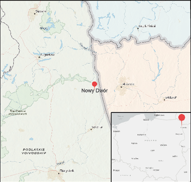
Being characterised by large, upright stones on graves, stelae cemeteries are a cultural phenomenon native to north-eastern Poland and Belarusian Grodno region (Fig. 1). Until recently, these necropolises have been associated with local Yotvingian communities, or – alternatively – interpreted as epidemic cemeteries. During the European late medieval and post-medieval period, it was a common, although not universal practice to establish epidemic cemeteries far from human settlements or sacred grounds. It should be noted, however, that in such cases the deceased were usually buried in mass graves, and the marking of their resting place was rarely permanent (Duma 2015: 142—143). Since in terms of funerary rite stelae cemeteries were often a necropolis with singular graves, thus, stelae cannot be interpreted as indicators of plague victims’ burials. On the other hand, the tradition of marking graves with stones or boulders is present in many local cultures – not only Yotvingian, but also Christian Orthodox, Uniate, Jewish and Muslim Tatar.
Fig. 1. Map with the location of Nowy Dwór. Created by Hubert Lepionka.
Since 2018, stelae necropoleis have been a subject of a research identification program led by the Podlasie Museum in Białystok, Poland. As part of the project, in 2020 the Podlasie Museum conducted rescue excavations in Nowy Dwór, where a previously unknown stelae cemetery was discovered during construction works. The study had two points: archaeological research and prospection, and anthropological analysis of unearthed remains. The following paper presents and discusses preliminary results of this study.
The exact date of the Nowy Dwór foundation is unknown and is, therefore, a subject of discussion. The earliest possible period was in the years 1440 and 1492 (Wiśniewski 2006: 149; Ryżewski 2006; 2019) or in 1505 (Ryżewski 2019: 124). During the time it was a royal property, Nowy Dwór became the centre of colonization and floatation of forest goods. In 1578 it was granted town privileges based on Magdeburg rights – an event that only sealed its status (Ryżewski 2006: 308—310).
The town population and its closest vicinity consisted mainly of colonizers coming from the east, from territories of the Grand Duchy of Lithuania (Wiśniewski 1978, Ryżewski 2006; 2019). In the first half of the 16th century, at the same time the Catholic Church of John the Baptist and an Orthodox church were founded in the town (Ryżewski 2019). With the Union of Brest, at the turn of the 16th century the Orthodox parish became Uniate.
Excavations in Nowy Dwór were conducted after the discovery of human remains and stones during an archaeological supervision of the reconstruction of a local road near the Orthodox church, and covered a 4-meter wide and 60-meter-long trench located on the North-Western border of the cemetery (Fig. 2). As a result, 88 graves typical of a Christian burial rite were discovered.
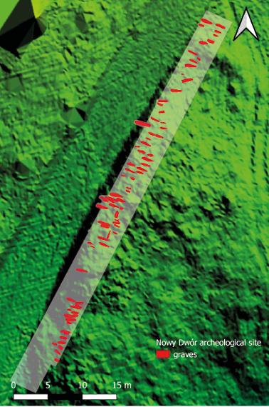
Fig. 2. Map of the site with graves. Created by Hubert Lepionka, drawn by Olga Dec.
On the excavated part of the cemetery 88 singular skeletal flat graves (occasionally with additional loose bones) were discovered, with the dead resting in extended supine position with either both upper limbs folded on the pelvis, or with the left hand over the heart area. In several graves remains of wooden caskets were found, although, they were not present with most burials, thus, leading to the conclusion that the deceased were buried primarily with shrouds. Similarly, barely any grave goods were discovered, and only in a few graves coins were present. Graves were mainly oriented from the North-West to South-East, with skulls pointing towards North-West. However, there were few deviations to this rule (Fig. 3). Only in two graves there was a completely different orientation: in the grave no. 75 the deceased was buried with their head to the South, and in the grave no. 73 to the South-West. Some of the graves were marked with a stone stelae with no visible traces of carvings or engravings. Preserved stelae were located in parts of the cemetery where the topsoil has not been previously disturbed (Fig. 4).
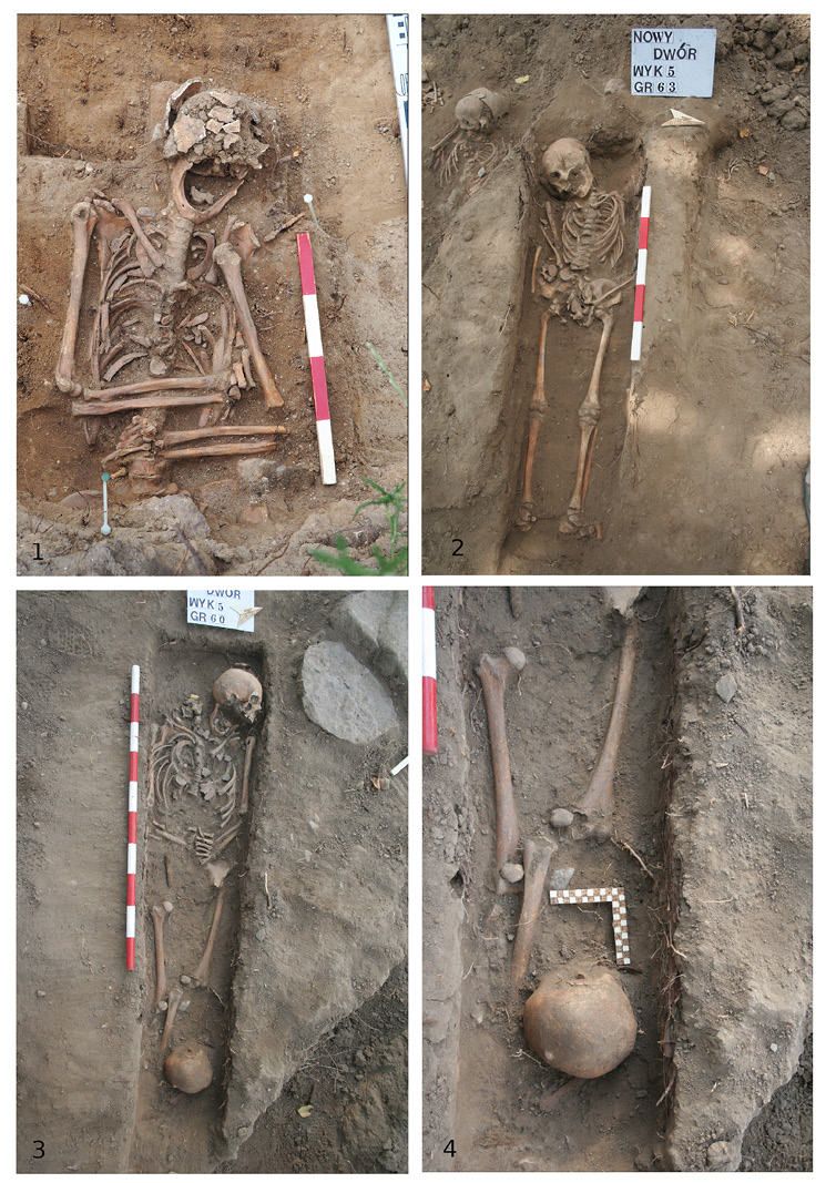
Fig. 3. Selected grave examples. (1) Grave no. 3, (2) Grave no. 63, (3) Grave no. 60, (4) Additional skull from the grave no. 60. Photographed by Hubert Lepionka.
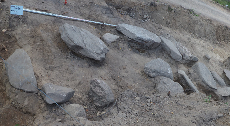
Fig. 4. Grave-marking stone stelae in the trench with an undisturbed top soil. Photographed by Hubert Lepionka.
In the surveyed section of the cemetery at least 181 people were buried. These graves had been intact by the time of discovery. The rest of the remains belonged to two different types of burials. First, there were features that can be described as consisting of small pits with a layer of loose bones of 2 to 3 individuals, who were buried in what resembled a pile. These were probably secondary burials of loose bones found on the surface of the cemetery (Fig. 5). The rest of bone material consisted of at least 60 individuals found in a mixed layer of various bones devoid of any anatomical arrangement upon discovery. It can be presumed that this may be related to the long-term intensive use of the cemetery, during which many graves were at least partially destroyed (Fig.6). Regardless of the condition of the in situ burials, no significant differences between people buried in regular graves and people from the mixed layer were observed. It is highly probable that original burials from this layer were identical to that of graves from other layers. Therefore, we treated all collected and analysed bones as one set.
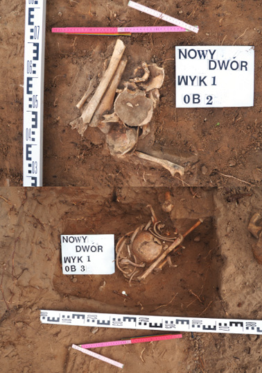
Fig. 5. Loose bone feature no. 2 (upper) and no. 3 (lower). Photographed by Hubert Lepionka.
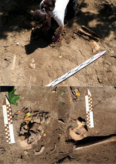
Fig. 6. Mixed layer during exploration. Photographed by Hubert Lepionka.
The sex of the adult individuals, whose remains were preserved in anatomical arrangement, was assessed based on the dimorphic features of the skull (Acsádi and Nemeskéri 1970) and pelvis (Buikstra and Ubelaker 1994). The pelvic bone was used for sex assessment for adult individuals belonging to the loose bones category. To sum up, the examined material included the remains of 65 children up to 15 years of age, 28 females, 36 males and 52 individuals of unspecified sex (tab. 1). The highest percentage of the remains of unknown sex derived from the loose bone material, by which the assessment of this parameter was insubstantial due to a limited number of pelvic bones. A second negative factor influencing sex assessment was the different state of bone preservation. This factor also contributed to the determination of the parameter of biological age at time of death. This characteristic in adult individuals was estimated based on morphological features of the surface of ribs’ sternal ends (Işcan et al. 1984), pubic symphysis (Todd 1921, Brooks and Suchey 1990), pelvic auricular surface (Lovejoy et al. 1985, Buckberry and Chamberlain 2002), and the phase of teeth wear (Lovejoy 1985).
Table 1. Age and sex estimation of individuals from post-medieval population of Nowy Dwór
| Age (years) |
Sex | N of indiv. in reg. graves |
% of indiv. in reg. graves |
N of indiv. in objects |
% of indiv. in objects |
N of indiv. in comingled layer |
% of indiv. in comingled layer |
| <0 | Undefined | 2 | 1.11 | 0 | 0.00 | 1 | 0.60 |
| 0-4 | Undefined | 23 | 12.71 | 2 | 1.11 | 20 | 11.05 |
| 5-9 | Undefined | 8 | 4.42 | 1 | 0.60 | 2 | 1.11 |
| 10-14 | Undefined | 4 | 2.21 | 0 | 0.00 | 2 | 1.11 |
| 15-19 | F | 0 | 0.00 | 0 | 0.00 | 2 | 1.11 |
| M | 3 | 1.66 | 0 | 0.00 | 0 | 0.00 | |
| NN | 6 | 3.32 | 1 | 0.60 | 0 | 0.00 | |
| 20-24 | F | 2 | 1.11 | 0 | 0.00 | 0 | 0.00 |
| M | 0 | 0.00 | 0 | 0.00 | 0 | 0.00 | |
| NN | 0 | 0.00 | 0 | 0.00 | 0 | 0.00 | |
| 25-29 | F | 1 | 0.60 | 0 | 0.00 | 0 | 0.00 |
| M | 2 | 1.11 | 0 | 0.00 | 0 | 0.00 | |
| NN | 0 | 0.00 | 0 | 0.00 | 0 | 0.00 | |
| 30-34 | F | 4 | 2.21 | 0 | 0.00 | 2 | 1.11 |
| M | 4 | 2.21 | 0 | 0.00 | 3 | 1.66 | |
| NN | 0 | 0.00 | 0 | 0.00 | 0 | 0.00 | |
| 35-39 | F | 0 | 0.00 | 0 | 0.00 | 0 | 0.00 |
| M | 3 | 1.66 | 0 | 0.00 | 2 | 1.11 | |
| NN | 0 | 0.00 | 0 | 0.00 | 0 | 0.00 | |
| 40-44 | F | 4 | 2.21 | 0 | 0.00 | 0 | 0.00 |
| M | 2 | 1.11 | 0 | 0.00 | 1 | 0.60 | |
| NN | 0 | 0.00 | 0 | 0.00 | 0 | 0.00 | |
| 45-49 | F | 1 | 0.60 | 0 | 0.00 | 0 | 0.00 |
| M | 4 | 2.21 | 0 | 0.00 | 0 | 0.00 | |
| NN | 0 | 0.00 | 0 | 0.00 | 0 | 0.00 | |
| 50-54 | F | 3 | 1.66 | 0 | 0.00 | 0 | 0.00 |
| M | 1 | 0.60 | 0 | 0.00 | 0 | 0.00 | |
| NN | 0 | 0.00 | 0 | 0.00 | 0 | 0.00 | |
| 55-59 | F | 0 | 0.00 | 0 | 0.00 | 0 | 0.00 |
| M | 0 | 0.00 | 0 | 0.00 | 1 | 0.60 | |
| NN | 0 | 0.00 | 0 | 0.00 | 0 | 0.00 | |
| 60+ | F | 2 | 1.11 | 0 | 0.00 | 0 | 0.00 |
| M | 4 | 2.21 | 0 | 0.00 | 0 | 0.00 | |
| NN | 1 | 0.60 | 0 | 0.00 | 0 | 0.00 | |
| NN | F | 6 | 3.32 | 1 | 0.60 | 0 | 0.00 |
| M | 5 | 2.76 | 1 | 0.60 | 0 | 0.00 | |
| NN | 16 | 8.84 | 2 | 1.11 | 26 | 9.39 | |
| Total | – | 111 | 61.56 | 8 | 9.04 | 62 | 29.45 |
| Total number | 181 | ||||||
| Total % | 100 | ||||||
In non-adult individuals, biological age at death was assessed by the stage of skeletal ossification and degree of development of primary and permanent teeth (Ubelaker 2018). Based on this data, examined individuals were assigned to one of the seven standard anthropological categories: fetus; infans I – up to 7 y.o.; infans II – up to 14 y.o.; juvenis – up to 22 y.o.; adultus – up to 35 y.o.; maturus – up to 55 y.o.; senilis – over 55 y.o. In the case of incomplete or badly preserved material, where the assessment of biological age at death was impossible, individuals were assigned to the child/adult category based on the development of preserved bones. This data, for the need of group and population analysis, was statistically evaluated between the anthropological age at death categories. To establish the demographic profile of the population, 14 age categories with 5-year ranges were created. Individuals were assigned to a specific category based on the features of biological age at death. Number of cases in certain categories is shown in table 1. Body height was calculated by measuring the length of the long bones (Trotter and Gleser 1952).
Among the analysed remains we recorded both sex variants, with a slight prevalence of male individuals (Table 1). However, this disproportion was probably due to the overall low percentage of adults with defined sex parameters. Remains in the surveyed area also represented all categories of age at time of death – from prenatal period to individuals over 60 years of age (Table 1). Based on the mortality tables constructed for the sample (Table 2) it is apparent that the highest number of deaths and one of the highest probabilities of death rates was recorded in the 0-4 years of age category. The second most numerous was 30-34 years of age category. The lowest number of deaths was observed in the 55-59 years of age category. Continued adult life expectancy for 20-year-old individuals – which is a measure of population quality – was approximately 22 years; meaning the average adult life expectancy of the population was 42 years. Similarly, the approximate percentage of life expectancy of 23 years was recorded for newborns.
Table 2. Mortality table for the post-medieval population of Nowy Dwór
| Age (in years) | Dx | dx | lx | qx | Lx | Tx | ex |
| 0-4 | 61 | 34.27 | 100.00 | 0.34 | 414.33 | 2 328.43 | 23.28 |
| 5-9 | 12 | 6.74 | 65.73 | 0.10 | 311.80 | 1 914.10 | 29.12 |
| 10-14 | 6 | 3.37 | 58.99 | 0.06 | 286.53 | 1 602.30 | 27.16 |
| 15-19 | 15 | 8.43 | 55.62 | 0.15 | 257.03 | 1 315.77 | 23.66 |
| 20-24 | 3 | 1.69 | 47.19 | 0.04 | 231.73 | 1058.74 | 22.44 |
| 25-29 | 5 | 2.81 | 45.50 | 0.06 | 220.48 | 827.01 | 18.18 |
| 30-34 | 23 | 12.92 | 42.69 | 0.30 | 181.15 | 606.53 | 14.21 |
| 35-39 | 9 | 5.06 | 29.77 | 0.17 | 136.20 | 425.38 | 14.29 |
| 40-44 | 13 | 7.30 | 24.71 | 0.30 | 105.30 | 289.18 | 11.70 |
| 45-49 | 9 | 5.06 | 17.41 | 0.29 | 74.40 | 183.88 | 10.56 |
| 50-54 | 7 | 3.93 | 12.35 | 0.32 | 51.93 | 109.48 | 8.87 |
| 55-59 | 2 | 1.12 | 8.42 | 0.13 | 39.30 | 57.55 | 6.84 |
| 60+ | 13 | 7.30 | 7.30 | 1.00 | 18.25 | 18.25 | 2.50 |
| Total | 178* | 100.00 | – | – | – | – | – |
* Remains of 37 adult individuals and 18 subadult individuals of undetermined precise age at time of death, were statistically evaluated. Remains of three individuals who died at prenatal age were not included in the mortality table.
Abbreviations:
Dx – number of individuals in x age (at time of death) category.
dx – percentage of individuals in x age (at time of death) category.
lx – percentage of individuals living to x years of age.
qx – probability of death at x years of age.
Lx – number of years lived overall by all individuals in the x age category.
Tx – number of years left to live for all individuals in the x age category.
ex – average life continuity for individuals in x age category.
Variation in body height was linked with determined sex. Average height was estimated at 159 centimetres for the females and 170 centimetres for males (Table 3).
Table 3. Height estimation of adult post-medieval population of Nowy Dwór
| Sex | Lowest height value (in cm) | Highest height value (in cm) | Average height (in cm) | SD | N |
| Female | 150 | 169 | 159 | 5,13 | 16 |
| Male | 160 | 180 | 170 | 4,66 | 23 |
Recorded markers of physiological stress on child remains, linear enamel hypoplasia from a mild to a moderate degree (acc. to Garcin 2010) was the most common and was present in approximately 33% of analysed subadults with preserved teeth. The second most numerous was cribra orbitalia, being from weak to moderate severity (acc. Steckel at al. 2006), which was observed in 25% of skeletons with preserved upper orbital edges. Lesions on the greater wings of sphenoid bones, posterior surfaces of maxillae, hard palate, in the form of porosity or periostitis occurrence were observed in approximately 12.5% of cases.
A similar pattern of physiological stress markers was observed within adult population. Additionally, dental caries of varying degrees of severity were recorded in approximately 66% of adult individuals, whereas with subadults these occurred only in a few cases. Apart from cribra orbitalia (12.5%), singular cases of cribra cranii were also reported. Porosities and periostitis around the base of skull and on the mandible, (probably due to vitamin C deficiency-related lesions in adults), were observed in 12.5% of cases. Furthermore, significant ante-mortem tooth loss and bone inflammations were common.
Lesions of an overload-degenerative nature, often associated with an active lifestyle, were among the most common in the analysed sample. Individuals aged 16-18 years of age at time of death were the youngest in whom musculoskeletal overuse was observed – based on severe ligamentum flavium ossification, Schmorl’s nodes on the vertebral bodies, and traces of osteochondritis dissecans in joints. However, it should be noted that only few juveniles were affected, and lesions of this type were common with older age groups, starting with young adults aged 20-29 years of age at the time of death. Enthesophytes on limb bones, vertebral osteophytes of varying severity, Schmorl’s nodes, ligamentum flavium ossification, and numerous cases of osteochondritis dissecansin were also observed in all individuals aged 20-29 years of age. All degenerative-related lesions – particularly in limb joints – in Nowy Dwór inhabitants increased both in severity and frequency in people over 45 years of age.
The examined material showed many cases of infection diseases as well. Based only on skeletons from intact graves, infection-related lesions were observed in 24% of cases. This percentage increased, however, if loose bone material was also included. Thus, it can be estimated that infection-related changes occurred in at least 33% of the population. One of the most frequently recorded indicators of possible infection was long bone periostitis in varying forms and severity (Fig. 7:1) – it was often present within a pair of limb bones (e.g. right and left tibia), but cases of single and multifocal periostitis involving different bones have also been reported. In several instances, these coexisted with lytic lesions, e.g. on the ribs or vertebrae. Periostitis was recorded twice as often in remains of children. Several examples of inflammatory reaction resulting in osteomyelitis of varying severity were also present, occurring in both singular bones, and larger parts of the entire skeleton (Fig. 7:2). Among both subadults and adults, inflammatory reaction of periosteum of varying severity and spread was also present, occurring on the inner surfaces of the cranial bones (Fig. 8:1).
Traces of infection were also been observed in the ribs, and can be divided into three categories. The first category, included cases of bone tissue superstructure on the inner surface of the rib shafts with uninterrupted tissue continuity (Fig. 8:2); the second category refered to local bone loss causing lytic lesions (Fig. 9:1); the third category included lytic lesions observed on the external surface of the ribs at the junction with thoracic vertebrae. Regardless of the type, these markers were present in both adult and subadult remains, and often co-occured with lytic lesions on vertebrae or with infection indicators on long bones. Lytic lesions on vertebrae could also be categorised according to the incidence location. The first category included local bone loss within vertebral bodies – these refer to singular incidences recorded only in adult remains. The second group consisted of lytic lesions within vertebral arch structure (Fig. 9:2), present in subadults.
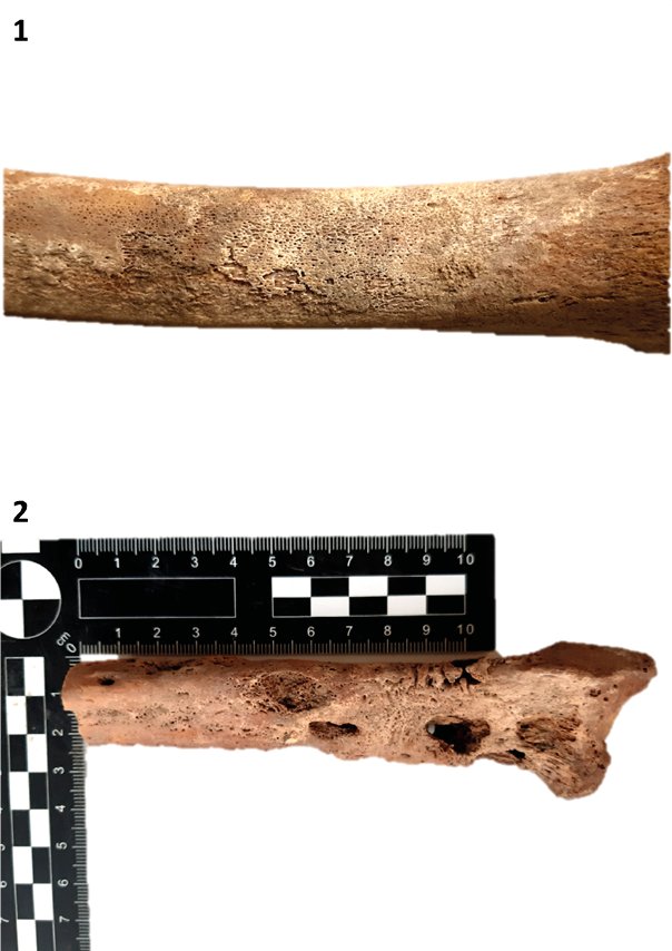
Fig. 7. Selected pathological condition examples. (1) Left tibial shaft with periosteal reaction, grave no. 70, (2) Distal fragment of left radius with advanced stage of Osteomyelitis, grave no. 22. Photographed by Angelika Słodka.
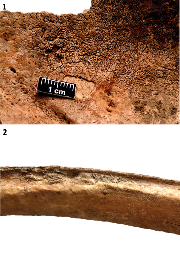
Fig. 8. Selected pathological condition examples. (1) Internal surface of occipital bone with periosteal reaction, loose bones, (2) Internal surface of rib with periosteal reaction, grave no. 53. Photographed by Angelika Słodka.
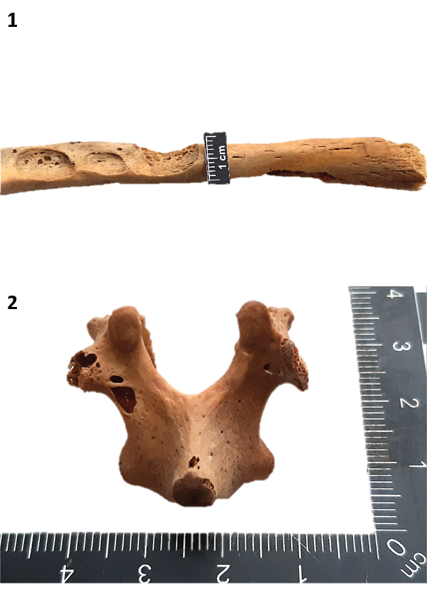
Fig. 9. Selected pathological condition examples. (1) Internal surface of rib with lytic lesions, grave no. 24, (2) Vertebral arch with lytic lesions, grave no. 57. Photographed by Angelika Słodka.
Furthermore, the repeatable occurrence of non-metric, epigenetic features in multiple individuals was also noted, among them numerous instances of cervical enamel projection in majority of examined teeth, repetitive patterns of Wormian bones, and several examples of non-obliterated metopic sutures. This in turn suggests probable high homogeneity and low gene flow within analysed population.
This study has showed that people were buried regardless of their biological profile within the surveyed part of the cemetary. What is puzzling, is the abundance of remains of children under 10 years of age at time of death, which accounts for 41% (after statistical evaluation) of all individuals. It is generally accepted that in most historical periods there was a high mortality rate in children, estimated at approximately 33% (Lewis 2007). In the Nowy Dwór case, this figure exceeds the generally accepted child mortaily rate. However, it should be noted that excavations covered only parts of the cemetery, and therefore, the examined sample represents only a fragment of entire population. It is possible that the observed high percentage of subadults may be related to the occurrence of additional environmental factors increasing the mortality, e.g. an epidemic, or location of child burials located near the end of the cemetery. For this reason, the estimated values correspond more to medieval than post-medieval populations (Kozłowski 2012; Pudło 2016; Budnik and Pudło 2017). Despite the significant underestimation of the parameters of the extinction table for a post-medieval population, the noted pattern does not characterise catastrophic extinction. What seems more likely was the occurrence of several events in short intervals, or a single long-term event. Confronting these values with historical data, it is possible that during the 18th century – and perhaps even earlier – events in Nowy Dwór significantly affected the town’s inhabitants, and thus, their life expectancy. In terms of body length, the analysed sample can be described as of middle height (Piontek 1992) for both females and males regardless of their age.
Several types of physiological stress were recorded. Disturbances in the enamel layer formation are usually associated with a general deficiency of minerals, proteins, or vitamins due to malnutrition. However, it cannot be excluded that linear enamel hypoplasia could form because of infection or various childhood diseases – therefore, it is considered a non-specific indicator (Irish et al. 2015). Cribra orbitalia formation is also not clear. In most cases it is associated with iron deficiency due to anaemia, yet among other possible causes are infectious diseases, parasites, or anaemias of genetic origin (Grauer 2011). Occurrences of local porosity and periostitits on the skull bones are lesions quite clearly identified as effects of scurvy and vitamin C deficiency; it should be taken into account that such deficiency may be due to both insufficient diet, as well as bodily need for vitamin C in case of infectious diseases (Halcrow et al. 2014). Lesions of overload-degenerative nature and their frequency are indicative of severe, long-time musculoskeletal overload most likely associated with physical labour from early adulthood.
An important task in material analysis is the verification of possible indicators of an epidemic mentioned in historical sources. Observations concerning the high mortality of children and mortality tables indicated the presence of an undefined factor that increased population mortality. Assessment of health and life conditions showed significant nutritional deficiencies among both subadults and adults, which impacted general immunity and in turn could have resulted in an epidemic increase. These elements, however, are not unequivocal indicators of an infectious disease in the population. For further verification, bone markers of infection – which were noted in up to 33% individuals and twice as often among children – were analysed. Periostitis is commonly regarded as an inflammatory lesion associated with infection, however, its nature does not point to any single pathogen that could be its cause. In addition, the issue of non-infectious factors (such as genetic, metabolic, and other diseases) have been raised (Weston 2012, Ortner 2003). Unlike periostitis, however, a relevantexample of an infection-related lesion is osteomyelitis. The most common aetiological agent associated with it is Staphylococcus aureus and Streptococcus causing purulent inflammation (Pinhasi and Mays 2008). Another possible pathogen is Mycobacterium tuberculosis. Several examples of inflammatory lesions of infectious origin occurred on the inner surfaces of the cranial bones. They were most likely results of a reaction of the periosteum – varying in severity and spread and noted in remains of both children and adults. Usually, they did not co-occur with other bone pathologies of infectious origin. It is probable that the placement of inflammatory lesions indicates its secondary nature and points towards infections of other systems, especially the respiratory system. Infections on the inner surfaces of the skull are often present in remains infected with Mycobacterium tuberculosis. Other pathogens (causing pneumonia) that are mentioned were equally as likely (Hershkovitz et al. 2002).
Traces of infection observed in the ribs can be divided into three categories. The most common cause of bone superstructure on ribs with uninterrupted tissue continuity are diseases associated with inflammation of the lower respiratory tract. On the other hand, cases of lytic lesions present at the rib-vertebral junctions are mostly attributed to tuberculosis (Davies-Barrett et al. 2019). Lytic lesions observed on the vertebrae are also categorised according to the place of their occurrence. These lesions are of varying degree of severity, progression and spread, and their occurrence – as with other infection markers – cannot be linked with one specific pathogen. However, changes of this type are most frequently present in cases of tuberculosis. Therefore, it may be possible, to some extent, to confirm historical reports on probable epidemics decimating a local population. Based on recorded and analysed pathologies, it is difficult to confirm their aetiological factor, since markers are most often non-specific. Nevertheless, it is worth noting that in the analysed material the most frequently recorded cases were those attributed to pulmonary infections and tuberculosis.
Based on available historical records, observed infection-related lesions can be associated with several time periods. The earliest information is one of the supposed demises of two post-Yotvingian villages near Nowy Dwór – Kopno and Jatwieź. The populations of both villages were believed to have died from plague at the end of 16th century (acc. to Żuk and Bujnowski 2009). Another period can be dated to the first half of 17th century, according to information included in letters of the servants of Radziwiłł family who had eyewitnessed the plague. The most reliable account derives from information that in 1706, 1707 and 1712 Nowy Dwór was first ravaged during the Northern War, and then (around 1760) affected by a plague which devastated almost the entire population, with only five families remaining (Ryżewski 2006: 316). It is possible that after the plague the population of the town was supplanted by the inhabitants of neighbouring villages.
Based on chronological findings, it was possible to trace the period of cemetery usage from 16th to half of the 18th century (Fig 10, 11). This closely corresponds with historical data from 1804, when inventory documentation of local Uniate parish was made, describing the church as being in bad condition at the time, as well as mentioning an old cemetery located next to the temple which was still in use. A new cemetery located on the peripheries of the town was also mentioned, however, it had not been in use during that time by the inhabitants. This suggests that the excavated area can be linked with the existence and functioning of the Orthodox and Uniate churches: beginning in the year 1530, and finally being abandoned in the first half of the 19th century. The abandonment of the cemetery also corresponds with the delegalisation of the Uniate religion by the decree of Tsar Nicholas I of Russia, and the foundation of a new Orthodox church next to where the previous temple was located (Ryżewski 2019).
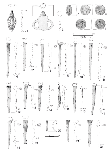
Fig. 10. Book binding piece (1), metal object (2), silver fragment of crucifix (3), copper coins of John II Casimir Vasa (4, 5) – loose finds from the layer between graves, coffin nails from graves (6–17). Drawn by Olga Dec.
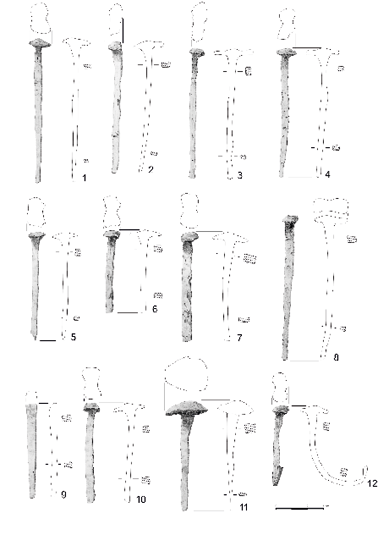
Fig. 11. Coffin nails from graves. Drawn by Olga Dec.
Among the findings, were a few numismatics which can be interpreted as “obols of the dead” – these are silver denars and two denars of Alexander and Sigismund II Jagiellon, minted in the period from early 16th to mid-16th century. Another set consists of small copper coins of John II Casimir Vasa, dated to the years 1659–1668, and used until the half of 18th century (Fig. 12). Several coins were found in the layers outside grave cavities, and most likely came from destroyed graves. The practice of the obol tradition can be divided into roughly two periods. First, it was from around the Early Middle Ages, then disappeared, and then emerged once again in the 14th century – only to reach its peak in the following centuries. This tradition was practiced in the regions the territory of modern Poland, Ukraine, Belarus, and Russia. In the case of the Polish-Ruthenian borderlands, it occurred mostly in rural areas. The cultural meaning and significance of the “obol of the dead” unclear. Some researchers raise the possibility of Arabic, Christian or even ancient influences, while others suggest probable fear for the fate of the deceased in the afterlife, or – alternatively – the fear of their return as malicious revenants (Miechowicz 2019).
Among the discovered graves some can additionally be described as “atypical” or “deviant” – i.e., as differing from the majority of graves in some respects. These included graves no. 75 and 73 – which were oriented differently from the rest, facing respectively South and South-West – as well as the grave marked with number 60 (Fig. 3:3,4). The latter is of particular interest, as it could have been subjected to unusual practices, exceptional in the scale of the examined parts of the necropolis.
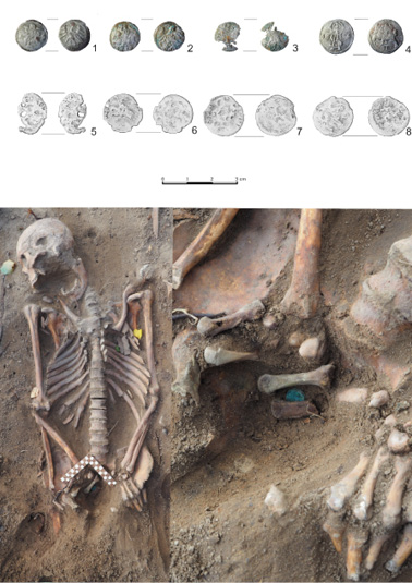
Fig. 12. “Obols of the dead”. Upper: (1) Coin of Alexander Jagiellon, 1501-1506, grave no. 56. (2) Coin of Sigismund II Augustus Jagiellon, 1559, grave no. 1. (3) Coin of Sigismund II August Jagiellon, 1560, grave no. 1. (4) Coin of Sigismund II Augustus Jagiellon 1566, grave no. 3. (5) Copper coin of John II Casimir Vasa, grave no. 43, (6–7) Copper coins of John II Casimir Vasa. Low: grave no. 30 with “obols” in situ. Photographed by Hubert Lepionka, drawn by Olga Dec.
Grave no. 60 can theoretically be considered a double grave, as it consisted of one mostly complete skeleton and an additional skull deposited near the skeleton’s legs. Upon discovery, the skeleton rested in an anatomical arrangement in extended supine position; despite its medium degree of completeness and imperfect state of preservation, it was possible to determine that the remains belonged to a person presenting female characteristics, who had died at the age of approximately 40–44 years of age. Further analysis did not reveal any peri-, ante- or postmortem injuries, however, pathologies in the form of loss of three molars, inflammation of the jawbone, and degenerative-strain lesions of the spinal bones and ribs were noted. The skull of the second individual – which was poorly preserved, also presented female characteristics and indicated adult age at time of death – was located between the legs of the skeleton.
The deviant forms of the burial in question is further emphasised by the fact that post-depositional processes and overlapping of two independent burials – an older and younger one – can be excluded with a fair amount of certainty. Both the skeleton and the lone additional skull were buried not only within the same burial cavity, but also in one coffin that bore no signs of post-depositional disturbances or partial destruction. No other graves, grave remains, bones, or other types of features were recorded in the immediate vicinity of the burial, ruling out the possibility of skull being moved unintentionally.
Furthermore, the position of the skull raises questions as well. As one of the determinants of burials of the revenants, it is generally associated with the belief in the undead rising from the grave to harm the living community. Alternatively, mostly in regard to Late Middle Ages, these burials can be interpreted as those of criminals and convicts. It should be noted, however, that in burials of the alleged revenants what the issue here is that the decapitated skeleton is from one person, and not – as it is in the case of grave no. 60 – an additional skull. It is possible that in this case we are in fact faced with a material trace of some local, unknown belief or resulting behaviours, but it is difficult to determine exactly what kind it would be: whether the grave is that of a revenant, or perhaps a whisperer (witch). There is the possibility of the grave was an ordinary double grave of two people buried together – one who had died earlier, and who later had their skull removed and placed in the coffin of the other. However, this does not explain the placement of the skull, a position clearly associated with the burials of those suspected of revenantism or of other forms of a supernatural nature. Yet, associating grave 60 with revenantism itself is unlikely. It may, however, be a burial of a szeptucha – a whisperer (whispess) or witch, as witchcraft activities are to this day practiced in the region. Information obtained from various sources from the local community indicate that in the past whisperers used bones collected from graves in rituals and for healing purposes. Thus, the additional skull could be a specific designation of the deceased, marking them as a witch.
The Nowy Dwór necropolis bears a number of features which characterised the health and socio-cultural elements of the community which used it. Firstly, based on population studies it is possible to explain the relative homogeneity and genetically undiversified character of local society. Pathological lesions, such as periostitis, osteomyelitis or lytic lesions, which were present in 33% of all examined individuals, seem to provide at least some argument based on historical accounts for several epidemic events which decimated the town’s population, and ultimately caused the entire settlement to regress into a village. The cemetery itself, however, is with all certainty not a specifically epidemic cemetery. The necropolis stands out from the rest of similar stelae burial sites due to its location within the settlement, and not along its peripheries. Based on numismatic finds and historical sources, it is possible to set the usage period of the cemetery between the 16th to mid-19th centuries. Moreover, it most probably functioned in connection with the now non-existent Uniate church built in 1530, the location of which was initially estimated based on geomagnetic and geo-radar surveys. However, further and more extensive research is required to verify the hypothesis concerning the church itself.
In general, results obtained from Nowy Dwór make a note-worthy contribution to the process of identification and characterisation of the whole phenomenon of stelae cemeteries and post-medieval period culture in North-Eastern Poland – providing a wider perspective on local traditions and memory, as well as cultural and religious diversity of the region.
Acknowledgements
This publication was financed by the Minister of Science and Higher Education (Grant No DNK/SP/463728/2020): Excellent Science – Support for scientific conferences. Funeralia Gnieźnieńskie – Man in the perspective of interdisciplinary research. Authors would like to thank the Municipal Office of Nowy Dwór for funding, and for making this project possible. Also, the local community of Nowy Dwór for providing not only information about the history of the town and its traditions, but also for their immense support and unfading interest throughout the research.
Conflict of interests
No conflict of interests was declared
Authors’ contributions
HL wrote introduction, used photographs, and fragments covering archaeological record and history of Nowy Dwór; AS did an anthropological analysis, statistical analysis, interpretation of results and wrote part of Results section; OD id an author of used drawings, Abstract, part of Results, Conclusions, and was responsible for translation entire manuscript to English.
Acsádi G, Nemeskéri J. 1970. History of Human Life Span and Mortality. Budapest: Akadémiai Kiadó.
Buckberry JL, Chamberlain AT. 2002. Age estimation from the auricular surface of the ilium: a revised method. Am J Phys Anthropol 119.3:231–39.
Bogin B. 1999. Patterns of human growth. Cambridge: Cambridge University Press.
Budnik A, Pudło A. 2017. Biodemografia nowożytnego Gdańska w świetle badań nad ossuariami. Możliwości rekonstrukcji i problemy metodyczne. In: Fontes Commentationesque ad res gestas gedani et pomeraniae 6. Gdańsk: Muzeum Archeologiczne w Gdańsku.
Buikstra JA, Ubelaker DH. 1994. Standards for datat collection from human skeletal remains. Fayetteville: Arkansas Archaeological Survey.
Brooks S, Suchey JM. 1990. Skeletal age determination based on the os pubis: a comparison of the Acsádi-Nemeskéri and Suchey-Brooks methods. Hum Evol 5(3):227–38.
Davies-Barrett AM, Antoine D, Roberts C. 2019. Inflammatory periosteal reaction on ribs associated with lower respiratory tract disease: A method for recording prevalence from sites with differing preservation. Am J Phys Anthropol 168:530–42.
Dec O. 2020. Potrzeba rekonceptualizacji wczesnośredniowiecznych pochówków “wampirów” z ziem polskich. Folia Praehistorica Posnaniensia 25:63–70.
Duma P. 2015. Śmierć nieczysta na Śląsku. Studia nad obrządkiem pogrzebowym społeczeństwa przedindustrialnego. Wrocław.
Garcin V, Veleminsky P, Trefny P, Alduc-Le Bagousse A, Lefebvre A, Bruzek J. 2010. Dental health and lifestyle in four early medieval juvenile populations: Comparisons between urban and rural individuals, and between coastal and inland settlements. Homo 61(6):421–39.
Grauer AL. 2011. A companion to paleopathology. Vol. 23. John Wiley & Sons.
Halcrow SE, Harris NJ, Beavan NJ, Buckley HR. 2014. First bioarchaeological evidence of probable scurvy in Southeast Asia: Multifactorial etiologies of vitamin C deficiency in a tropical environment. Int J Paleopathol 5:63–71.
Hershkovitz I, Greenwald CM, Latimer B, Jellema LM., Wish-Baratz S, Eshed V. 2002. Serpens Endocrania Symmetrica (SES): A New Term and a Possible Clue for Identifying Intrathoracic Disease in Skeletal Populations. Am J Phys Anthropol 118:201–6.
Irish JD, Scott GR. 2015. A companion to dental anthropology. John Wiley & Sons.
Işcan MY, Loth SR, Wright RK. 1984. Age estimation from the rib by phase analysis: White females. J Forensic Sci 29:1094–104.
Işcan MY, Loth SR, Wright RK. 1985. Age estimation from the rib by phase analysis: White males. J Forensic Sci 30:853–63.
Kozak Ł. 2021. Upiór. 2nd edition. Warsaw.
Kozłowski T. 2012. Stan biologiczny i warunki życia ludności in Culmine na Pomorzu Nadwiślańskim (X–XIII wiek). Studium antropologiczne. Toruń: Wydawnictwo naukowe UMK.
Lepionka H, Cmentarzyska ze stelami a praktyki pogrzebowe chłopów litewskich w okresie nowożytnym. Małe Miasta Duchowość Kanoniczna, 581–97.
Lepionka H, Rybska M. 2017. Cmentarzysko ze stelami kamiennymi w Jagintach gm. Nowy Dwór, woj. podlaskie w świetle badań nieinwazyjnych, Podlaskie Zeszyty Archeologiczne, 201–10.
Lepionka H, Słodka A, Ryżewski G, Drupka B. 2020. Cmentarzysko ze stelami kamiennymi w Jagintach, gm. Nowy Dwór, woj. podlaskie w świetle badań interdyscyplinarnych. Podlaskie Zeszyty Archeologiczne, 139–55.
Lewis ME. 2007. The Bioarcheology of Children. Perspectives from Biological and forensic anthropology. Vol. 50. Cambridge University Press.
Lovejoy CO, Meindl RS, Pryzbeck TR, Mensforth RP. 1985. Chronological matamorpgosis of the auricular surface of the ilium: a new method for the determination of adult skeletal age at death. J Forensic Sci 68(1):15–28.
Lovejoy CO. 1985. Dental wear in the Libben population: its functional pattern and role in the determination of adult skeletal age at death. J Forensic Sci 68(1):47–56.
Łożyński K. 2006. Początek kolonizacji Puszczy Grodzieńskiej. In: Śliwiński J, editor.
Ortner DJ. 2003. Identification of pathological conditions in human skeletal remains. J Clin Forensic Med 13:154.
Pinhasi R, Mays S. 2008. Advances in human paleopathology. John Wiley & Sons.
Puszcze wielkoksiążęce na północnym Podlasiu i zachodniej Grodzieńszczyźnie w XV–XVI wieku (podziały, administracja, służby leśne i wodne). Olsztyn, 149.
Piontek J. 1992. Stres w populacjach pradziejowych. Założenia, metody, wstępne wyniki badań. Biologia Populacji Ludzkich Współczesnych i Pradziejowych. Słupsk, 321–345.
Pudło A. 2016. Mieszkańcy średniowiecznego Gdańska w świetle wyników badań antropologicznych. Fontes Xommentationesque ad Res Gestas Gedani et Pomeraniae 5. Gdańsk: Muzeum Archeologiczne w Gdańsku.
Sierba M. 2016. Morowe powietrze w Orli, na Podlasiu i w Rzeczpospolitej w listach urzędników podlaskich Krzysztofa II Radziwiłła – Macieja Berzeńskiego i Stanisława Kurosza. Studia Podlaskie 24: 41–59.
Stachowski K. 2005. Wampir na rozdrożach. Etymologia wyrazu upiór ~ wampir w językach słowiańskich. Rocznik Slawistyczny 55:73–92.
Steckel RH, Sciulli PW, Larsen CS, Walker PL. 2006. The Global History of Health: Data Collection Codebook.
Todd TW. 1921. Age changes in the pubic bone. Am J Phys Anthropol 4(1):1–70.
Trotter M, Glesser GC. 1952. Estimation of stature from long bones of American Whites and Negros. Am J Phys Anthropol 10(4):463–514.
Ubelaker DH. 2018. Estimation of Immature Age From the Dentition. New Perspectives in Forensic Human Skeletal Identification. Academic Press, 61–64.
Weston DA. 2012. Nonspecific infection in paleopathology: interpreting periosteal reactions. In: AL Grauer, editor. A Companion to Paleopathology. Oxford, UK: Wiley-Blackwell.
Wiśniewski J. 1967. Dzieje osadnictwa w pow. augustowskim od XV do końca XVIII wieku. In: Studia i materiały do dziejów Pojezierza Augustowskiego, Białystok 1967:55.


Received: 2021.10.11; Revised: 2022.05.12; Accepted: 2022.05.19