Available online at: https://doi.org/10.18778/1898-6773.85.3.06
Institute of Biological Sciences, Cardinal Stefan Wyszyński University in Warsaw, Wóycickiego 1/3, building 23, 01-938 Warsaw
Institute of Biological Sciences, Cardinal Stefan Wyszyński University in Warsaw, Wóycickiego 1/3, building 23, 01-938 Warsaw
Institute of Biological Sciences, Cardinal Stefan Wyszyński University in Warsaw, Wóycickiego 1/3, building 23, 01-938 Warsaw
Institute of Biological Sciences, Cardinal Stefan Wyszyński University in Warsaw, Wóycickiego 1/3, building 23, 01-938 Warsaw
Institute of Biological Sciences, Cardinal Stefan Wyszyński University in Warsaw, Wóycickiego 1/3, building 23, 01-938 Warsaw
Institute of Biological Sciences, Cardinal Stefan Wyszyński University in Warsaw, Wóycickiego 1/3, building 23, 01-938 Warsaw
ABSTRACT: The aim of this analized is to evaluate the frequency of osteoarthritis in the early modern population of Dąbrówki (Poland). Evaluation of degenerative joint changes was based on standard methods commonly used in physical anthropology. Three types of changes were studied: osteophytes, porosities, and eburnations. They were analyzed in the shoulder, elbow, wrist, hip, knee, and proximal ankle joints. Osteoarthritic changes were assessed in 24 female, 20 male, and 8 undetermined sex individuals in the Dąbrówki population.
In the population from Dąbrówki the highest frequency of degenerative changes was noted in the hip joint, and the lowest in the knee joint. Osteophytes were the predominant type of lesions. The less frequent type was porosity, while polishing of the articular surfaces did not occur. In males, degenerative changes were noted more frequently than in females. Due to the existence of many interpretative limitations (there is no a complete picture of the population from Dąbrówki - skeletal material under exploration; not entirely clear and multifactorial etiology of degenerative joint changes), further analysis of the markers of environmental stress in the population from Dąbrówki is necessary.
KEY WORDS: osteoarthritis, skeletal population, osteophytes, porosity, eburnation
Osteoarthritis (OA) is the most ubiquitous pathological condition in skeletal populations (Ortner, Putschar 1985; Weiss, Jurmain 2007). It is also the most common joint condition observed today (Arden, Nevitt 2006; Rothschild, Woods 2012).
The aetiology of osteoarthritis is multifactorial (Felson 2003; Gabay et al. 2008; Roach and Tilley 2007; Teichtahl et al. 2005). In the clinical literature, an influence of several main factors has been considered. Age, sex, genes, metabolic and endocrine factors, obesity, bone density, articular cartilage nutrition, joint injuries and infection, joint instability, congenital and/or developmental joint deformities, physical activity and occupation, muscle weakness (Anderson, Loeser 2010; Arden, Nevitt 2006; Felson 2003; Gabay et al. 2008; Teichtahl et al. 2005; Guan et al. 2019), or even nutritional factors (Guan et al. 2019; Tetlow and Woolley 2001) are considered.
Given a high prevalence and availability of osteoarthritis in skeletal material (Weiss, Jurmain 2007; Plomp et al. 2015), the analyses of these skeletal markers of environmental stress have an important place in anthropology (Weiss, Jurmain 2007; Jurmain 1991; Klaus et al. 2009; Molnar et al. 2011; Schrader 2012; Woo, Pak 2013; Rojas-Sepúlveda, Dutour 2014; Palmer et al. 2016).
In spite of the fact that osteoarthritic changes are very common in historical populations, these pathological traits still raise many methodological and interpretative problems (Weiss, Jurmain 2007). The present study aimed to examine the osteoarthritic changes in the early modern skeletal material from Dąbrówki (Poland), the OA changes presence and the way they are expressed. It enriches the knowledge about these conditions in past skeletal populations, what is important in a reliable interpretation of the biology, ecology, and behaviour of past human populations.
All analysed specimens were excavated from a cemetery located in Dąbrówki (Poland). „Dąbrówki” as the name first appeared in 1559 (due to the presence of oaks in the local part of Podlasie). In 1568, the village of Dąbrówki was mentioned when the parish near Wasilków was established (Wawrzeniuk 2021a).
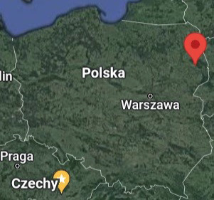
Fot. 1. The location of the Dąbrówki (red drop).
The cemetery with skeleton burials was discovered by accident in 2018, during earthworks. The cemetery on the hill is locally called „Brama Cygańska”. However, it is not known where this name comes from. There is also no information about the cemetery itself (Wawrzeniuk 2021a). With the help of the found burial equipment, the chronology of the cemetery can be estimated. The coins from the times of Sigismund Augustus and Sigismund III Vasa, a metal cross, a copper ring and a rosary bead, may indicate that this part of the cemetery under study dates back to the beginning of the 17th century. It cannot be ruled out that in the remaining areas of the cemetery there are burials from younger and older periods (Wawrzeniuk 2021b).
Numerous human remains were found on the hillside (Wawrzeniuk 2021a). Research work has revealed 62 graves (41 have been explored, 21 in total). Most of the individuals were adults. There were few children’s burials and they were usually buried with adults (Wawrzeniuk 2021a; Wawrzeniuk 2021b).
The samples evaluated for this analysis is a part of the osteological collection of the Institute of Biological Sciences Cardinal Stefan Wyszynski University (Poland). The exact number of male and female individuals in each historical group examined in this study is presented in Table 1.
Standard anthropological methods were applied to determine the age and sex of the individuals. The sex of the individuals was estimated using the Phenice (1969) and Buikstra, Ubelaker (1994). This includes visual assessments of pelvic and cranial features. The age at death of the individual was estimated based on changes in the morphology of the pubic symphysis, using the Brooks and Suchey (1990) system and standards for changes in the topography of the auricular surface (Buikstra, Ubelaker 1994). Only adult remains were included in this study. Individuals without any observable (except from osteoarthritic) skeletal changes (like observable bone illnesses, traumas, fractures, or bone deformities) were included into the analyses.
Osteoarthritic changes were examined in accordance with the methods by Buikstra and Ubelaker (1994). Three types of OA changes were examined: [a] osteophytes (OP) – a marginal proliferation of new bone in either the horizontal or vertical direction that produces a change in the shape of the joint contour; [b] porosity (POR) – pitting and/or erosion of the joint surface; [c] eburnation (EB) – polished subchondral bone with or without ridges) (Buikstra & Ubelaker 1994). OA changes were scored in: [a] shoulder (articular surface of the scapula and humeral head); [b] elbow (articular surfaces of the distal end of the humerus and articular surfaces of the proximal end of the ulna and radius); [c] wrist (articular surfaces of the distal end of the ulna and articular surfaces of the distal end of the radius); [d] hip (acetabulum [pelvic bone] and articular surface of femoral head); [e] knee (articular surface of the distal end of the femur and articular surfaces of the proximal end of the tibia); and [f] ankle (distal end of the tibia). Due to the small size of the sample, statistical analyzes did not include the gradation of changes, but only their presence (1) or their absence (0). The data on degenerative changes were analyzed separately for female, male and indeterminate sex, by joint analysis.
The statistical significance of the differences in the frequency of the degenerative changes between males and females in the analyzed skeletal material was investigated using the unilateral test for two components of the structure. The test analyses the significance of the differences between the frequencies of the two compared traits. Statistical significance was determined at the level of p≤0.05. All analyses were done using Statistica 13.3.
In the population from Dąbrówki, degenerative changes in joints were reported in 81% of individuals. The highest rates of degenerative changes were recorded in the hip joint (55%). Lower rates were recorded in the following joints: wrist (40%), shoulder (40%), elbow (40%), ankle (33%) and knee (27%). In the material studied, osteophytes were the predominant type of lesions (59%). The less frequent type of lesions was porosity (5.5%). There was no sanding of the articular surfaces (Tab. 1).
In the examined skeletal material, osteophytes were the most frequently observed degenerative changes in the knee (67%) and elbow (63%). Fewer osteophytes were observed in the shoulder (60%), hip (57%), wrist (57%) and ankle (55%) joints, respectively. The highest porosity was recorded in the hip joint (21%). Less in the shoulder (5%), elbow (2%) and knee (1%) joints. There was no porosity in the wrist and ankle joints (Tab. 1).
| Joint | N/n (% n) | |||
| Osteophytes | Porosity | Eburnation | All changes | |
| Shoulder | 45/27 (60,0%) | 62/3 (4,84%) | 63/0 (0,00%) | 67/27 (40,3%) |
| Elbow | 46/29 (63,04%) | 61/1 (1,64%) | 61/0 (0,00%) | 72/29 (40,28%) |
| Wrist | 37/21 (56,76%) | 52/0 (0,00%) | 52/0 (0,00%) | 52/21 (40,38%) |
| Hip | 72/41 (56,94%) | 81/17 (20,99%) | 83/0 (0,00%) | 83/46 (55,42%) |
| Knee | 30/20 (66,67%) | 68/1 (1,47%) | 72/0 (0,00%) | 75/20 (26,67%) |
| Ankle | 44/24 (54,55%) | 73/0 (0,00%) | 73/0 (0,00%) | 73/24 (32,88%) |
| All joints | 274/162 (59,12%) | 397/22 (5,54%) | 404/0 (0,00%) | 422/167 (39,57%) |
N – numer of the examined joints, n – number of joints with registered osteoarthritic changes
In females, degenerative changes in joints were reported in 79% of individuals. The highest number of changes of the osteophyte type was recorded in the ankle joint (75%). Less occurred in the knee (70%), wrist (63%), elbow (57%), hip (53%) and hip (45%) joints. Porosity occurred only in the hip joint with a frequency of 9% (Tab. 2). In males, degenerative changes were reported with a frequency of 100%. The greatest number of osteophytes was found in the shoulder joint (71%). Slightly less in the following joints: elbow (67%), knee (65%) and hip (59%). The lowest rates were recorded in the wrist (52%) and ankle (37%). Porosities occurred only in the joints: hip (36%), shoulder (12%) and elbow (3%) (Tab. 2). In the group of skeletons of undefined sex, the frequency of degenerative changes was 38%. Osteophytes, reported with a frequency of 100%, occurred in four joints (shoulder, elbow, hip and ankle). They occurred in the knee joint with a frequency of 67%. However, no osteophytes were found in the wrist joint. Porosity occurred only in the knee joint (14%) (Tab. 2).
| Joint | ||||||||||||
| Males | Females | Undefined sex | ||||||||||
| OP | Porosity | EB | All changes | OP | Porosity | EB | All changes | OP | Porosity | EB | All changes | |
| Shoulder | 20/9 (45%) |
33/0 (0,00%) |
32/0 (0,00%) |
34/9 (26,47%) |
24/17 (70,83%) |
25/3 (12%) |
27/0 (0,00%) |
29/17 (58,62%) |
1/1 (100%) |
4/0 (0,00%) |
4/0 (0,00%) |
4/1 (25%) |
| Elbow | 21/12 (57,14%) |
27/0 (0,00%) |
27/0 (0,00%) |
38/12 (31,58%) |
24/16 (66,67%) |
30/1 (3,33%) |
30/0 (0,00%) |
30/16 (53,33%) |
1/1 (100%) |
4/0 (0,00%) |
4/0 (0,00%) |
4/1 (25%) |
| Wrist | 16/10 (62,5%) |
25/0 (0,00%) |
25/0 (0,00%) |
25/10 (40%) |
21/11 (52,38%) |
25/0 (0,00%) |
25/0 (0,00%) |
25/11 (44%) |
0/0 (0,00%) |
2/0 (0,00%) |
2/0 (0,00%) |
2/0 (0,00%) |
| Hip | 38/20 (52,63%) |
43/4 (9,3%) |
44/0 (0,00%) |
44/21 (47,73%) |
32/19 (59,38%) |
36/13 (36,11%) |
36/0 (0,00%) |
36/23 (63,89%) |
2/2 (100%) |
2/0 (0,00%) |
3/0 (0,00%) |
3/2 (66,67%) |
| Knee | 10/7 (70%) |
28/0 (0,00%) |
29/0 (0,00%) |
31/7 (22,58%) |
17/11 (64,71%) |
33/0 (0,00%) |
34/0 (0,00%) |
35/11 (31,43%) |
3/2 (66,67%) |
7/1 (14,29%) |
9/0 (0,00%) |
9/2 (22,22%) |
| Ankle | 12/9 (75%) |
32/0 (0,00%) |
32/0 (0,00%) |
32/9 (28,13%) |
27/10 (37,04%) |
30/0 (0,00%) |
30/0 (0,00%) |
30/10 (33,33%) |
5/5 (100%) |
11/0 (0,00%) |
11/0 (0,00%) |
11/5 (45,45%) |
| All joints |
117/67 (57,26%) |
188/4 (2,13%) |
189/0 (0,00%) |
204/68 (33,33%) |
145/84 (57,93%) |
179/17 (9,5%) |
182/0 (0,00%) |
185/88 (47,57%) |
12/11 (91,67%) |
30/1 (3,33%) |
33/0 (0,00%) |
33/11 (33,33%) |
N – numer of the examined joints, n – number of joints with registered osteoarthritic changes, OP – osteophytes, EB – eburnation
When examining the significance of differences between males and females in the rates of degenerative changes in joints (all types of changes jointly), it was found that in men degenerative changes were recorded more often than in females (33% – females, 48% – males, p = 0.001). They were observed more often in men in each of the examined joints, with significantly higher rates reported for the shoulder joint (27% – females, 59% – males, p = 0.005) and elbow (32% – females, 53% – males, p = 0.005). = 0.04) (Tab. 3).
| Joint | Osteophytes | Porosity | Eburnation | All changes | ||||
| M/F | p | M/F | p | M/F | p | M/F | p | |
| Shoulder | 45/71% | 0,04* | 0/12% | 0,021* | 0/0% | – | 27/59% | 0,005* |
| Elbow | 57/67% | 0,251 | 0/3% | 0,182 | 0/0% | – | 32/53% | 0,040* |
| Wrist | 63/52% | 0,252 | 0/0% | 1,0 | 0/0% | – | 40/44% | 0,387 |
| Hip | 53/59% | 0,307 | 9/36% | 0,002* | 0/0% | – | 48/64% | 0,076 |
| Knee | 70/65% | 0,395 | 0/0% | 1,0 | 0/0% | – | 23/31% | 0,233 |
| Ankle | 75/37% | 0,026* | 0/0% | 1,0 | 0/0% | – | 28/33% | 0,335 |
| All joints | 57/58% | 0,436 | 2/10% | 0,001* | 0/0% | – | 33/48% | 0,001* |
p – statistically significant differences at the level of p≤0.05; F – frequency of degenerative changes in females, M – frequency of degenerative changes in males
The frequencies of osteophytes were similar in both sexes (57% – females, 58% – men). When examining the difference in the frequency of osteophytes in individual joints, significantly higher frequencies of these changes were noted in men in the shoulder joint (45% - females, 71% – males, p = 0.04), and in females in the ankle joint (75% – females, 37% – males, p = 0.04). In the wrist and knee joints, osteophytes were more common in females, in the elbow and hip joints – in males, however, these differences are not statistically significant (Table 3).
Porosity of the articular surface was noted more often in men in the shoulder joint (0% – females, 12% – males, p = 0.021), hip (9% – females, 36% – males, p = 0.002) and when all joints were analyzed jointly (2% – females, 10% – males, p = 0.001) (Tab. 3).
In the population from Dąbrówki, degenerative changes in joints were reported in 81% of individuals. The highest rates of degenerative changes were recorded in the hip joint (55%). Lower rates were recorded in the following joints: wrist (40%), shoulder (40%), elbow (40%), ankle (33%) and knee (27%). In the material studied, osteophytes were the predominant type of lesions (59%). The less common type of lesions was porosity (6%). There was no sanding of the articular surfaces (Tab. 1, Fot. 1).
In this population, degenerative joint changes were reported more often in men (48%) than in females (33%) (Table 3). They were observed more often in males in each of the examined joints, with significantly higher rates reported for the shoulder (27% – females, 59% – males) and elbow (32% – females, 53% – males) (Tab. 3). The frequencies of osteophytes were similar in both sexes (57% – females, 58% – males). When examining the difference in the frequency of osteophytes in individual joints, significantly higher frequencies of the changes were noted for males in the shoulder joint (45% – females, 71% – males), and for females in the ankle joint (75% – females, 37% – males). The porosity of the articular surface was noted more often in males in the shoulder (0% – females, 12% – males), in the hip (9% – females, 36% – males) and when all joints were analyzed together (2% for females, 10% for males) (Table 3).
The present study results are similar to other anthropological data (Weiss, Jurmain 2007). In majority of studies males were usually more affected than females (Šlaus 2002; Klaus et al. 2009; Eng 2016). In some skeletal materials, females (or some joints in females) have higher frequencies of OA than males (Molnar et al. 2011; Eng 2016). Some researchers did not find any significant (or very small) sex differences in OA (Eshed et al. 2010; Schrader 2012; Woo, Pak 2013; Palmer et al. 2016). Taking that osteoarthritic changes are treated by some researchers as physical activity markers (Capasso et al. 1999; Kennedy 1989), the obtain differences (or its lack) were connected with differences/similarities in lifestyle between sexes. Taking the above, it could be hypothesised that the sex differences in osteoarthritis in Dąbrówki population are a result of differences in daily activity patterns. Certain limitations dictate caution in drawing final conclusions. Firstly, archaeological, historical, and anthropological data about population from Dąbrówki is poor now, therefore at this stage of the studies it is not possible to say something about health, lifestyle of each individual. Finding out the relationships between osteoarthritic changes and physical activity is possible now and further studies are needed. Secondly, some researchers dictate caution in using OA changes as physical activity markers (e.g. Schrader 2012; Woo, Pak 2013), emphasizing mutifactorial etiology of these skeletal features. Finally, specifics of the skeleton material (usually small sample size, not well preserved bones, problems with sex determination) could not be omitted. It inflences the obtained results and its interpretation.
The skeleton material from Dąbrówki slightly differs in terms of the frequency of degenerative changes from other historical populations in Poland. In the population from Cedynia (14th–17th century) (Malinowska-Łazarczyk 1982; Porzeziński 2006), degenerative changes occurred with a frequency of 36% in men and 29% in females. The most common type of changes in both females and men were osteophytes. The joint with the most degenerative changes was the elbow joint (Myszka 2016).
In the modern population of Radom (17th-19th century), degenerative changes were reported with a frequency of 26% in the group of men and in 27% of females. The greatest number of lesions was observed in the hip (46%) and elbow (42%). Osteophytes were present in 24% of cases and it was the most frequently reported type of lesions (Myszka 2022).
In the population from Słaboszewo (14th–19th century) (Piontek 1977), degenerative changes occurred with a similar frequency in both sexes (24% in men and 25% in females). The most common type of changes in both sexes were osteophytes, and the joint where degenerative changes occurred most often - also regardless of sex – was the hip joint (Myszka 2016).
In the population from Łekno (14th–16th century) (Wyrwa 2003), degenerative changes occurred with a frequency of 38% in males and 32% in females. The most common type of lesions, as well as in the above-mentioned populations, were osteophytes. In males, degenerative changes were most often found in the elbow joint, while in females – in the hip joint (Myszka 2016).
Compared to the above-mentioned populations, degenerative changes in the population from Dąbrówki were more common in males (Dąbrówki 48%, other populations 24%–38%). In females, however, this difference was not significantly higher. Another point that distinguishes the population from Dąbrówek is the lack of polishing of the pond surfaces. Although in other populations they were rare, they were nevertheless recorded. Of particular interest is the fact that in the population from Dąbrówki, porosity in the hip joint was much more common in males (36%) than in females (9%), which significantly differs from the results of other populations (Myszka 2016).
Given the inadequate etiology of the degenerative changes, it is difficult to explain the cause of these discrepancies. Taking into account the fact that degenerative changes in joints are considered by some researchers as markers of physical activity (Capasso et al. 1999; Kennedy 1989), it can be assumed that one of the possibilities of explaining this phenomenon may be, different from other populations, the motor activity of individuals from Dąbrówek. However, nothing can be said about the physical activity of this population, because there is insufficient historical and archaeological data.
The obtained inter-population differences can be also a result of the differences between the type of settlement complexes. Cedynia (Zachodniopomorskie) was functioning as an early town settlement (Malinowska-Łazarczyk 1982). Łekno (Wielkopolska) was a part of the Łekno settlement complex where in historical times settlements and architectural structures of considerable political, administrative, socio-economic and religious significance were located (Wyrwa 1989). Population from Radom was characterised as a town population (Tomczyk et al. 2017). The population of Słaboszewo (Kujawsko-Pomorskie) represented a typical local rural society (Piontek 1981). Differences in technological development, architectural structures of the complexes, their political, administrative, socio-economic and religious significance influenced the socio-economic position of each populations and thus its health status, and biology. Different localization and not entirely convergent chronology of the compared archaeological series could also play a role in the obtained results. Another factor influencing the observed discrepancies may be the size of the sample, which is not large in the population from Dąbrówki.
Further studies of degenerative changes in the population from Dąbrówek are necessary, inter alia, to complete the knowledge about the population itself and to enrich knowledge about degenerative changes in joints in old skeletal populations, which due to the not fully understood etiology of changes and difficulties interpretative is of great importance.
In the population from Dąbrówki, degenerative changes in joints were reported in 81% of individuals. The most frequently reported type of lesions were osteophytes (59%). Porosity was noted with a frequency of 6%. Eburnation was not observed here. The highest rates of degenerative changes were recorded in the hip joint (55%). Lower rates were recorded in the following joints: wrist (40%), shoulder (40%), elbow (40%), ankle (33%) and knee (27%). In this population, degenerative changes in joints were reported more often in males (48%) than in females (33%). They were observed more often in males in each of the examined joints, with significantly higher rates reported for elbow and shoulder. The frequencies of osteophytes were similar in both sexes. Taking individual joint osteophytes were more frequently observed for males in the shoulder, for females in the ankle. The porosity of the articular surface was noted more often in males with higher frequencies in shoulder, and hip.
Due to the existence of many limitations resulting from the fact that the skeletal material is being explored and the etiology of degenerative changes in joints is not entirely clear and multifactorial, it is necessary to further analyze these determinants of environmental stress in the population from Dąbrówki.
Conflict of interests
Authors declared no conflict of interests.
Authors’ contribution
AK – carrying out skeletal analyses, preparation and description of the statistical part; JP – carrying out skeletal analyses, preparation and description of the statistical part; JW – preparation and description of the archaeological part; JT – content supervision; ZW – helping in statistical analysis; AM – planning and supervision of the research, setting a goal, substantive supervision, corresponding author.
Anderson AS, Loeser RF. 2010 Why is osteoarthritis an age-related disease? Best Practice & Research Clinical Rheumatology 24(1):15–26, https://doi.org/10.1016/j.berh.2009.08.006
Arden N, Nevitt MC. 2006. Osteoarthritis: epidemiology. Best Practice & Research Clinical Rheumatolog 20(1):3–25, https://doi.org/10.1016/j.berh.2005.09.007
Brooks S, Suchey JM. 1990. Skeletal age determination based on the os pubis: A comparison of the Acsádi-Nemeskéri and Suchey-Brooks methods, https://doi.org/10.1007/BF02437238
Buikstra JE, Ubelaker DH. 1994. Standards for data collection from human skeletal remains.
Capasso L, Kennedy KAR, Wilczak CA. 1999. Atlas of Occupational Markers on Human Remains, Journal of Paleontology, Monographic Publication, 3, Edigrafital S.p.A., Teramo.
Eng JT. 2016. A bioarchaeological study of osteoarthritis among populations of northern China and Mongolia during the Bronze Age to Iron Age transition to nomadic pastoralism. Quaternary International 405:172–175, https://doi.org/10.1016/j.quaint.2015.07.072
Eshed V, Gopher A, Pinhasi R, Hershkovitz I. 2010. Paleopathology and the origin of agriculture in the Levant. American Journal of Physical Anthropology 143(1):121–133, https://doi.org/10.1002/ajpa.21301
Felson DT. 2003. Epidemiology and osteoarthritis in: Osteoarthritis. Second edition. Oxford: Oxford Medical Publications 9–16.
Gabay O, Hall DJ, Berenbaum F, Henrotin Y, Sanchez C. 2008. Osteoarthritis and obesity: experimental models. Joint Bone Spine 75(6):675–679, https://doi.org/10.1016/j.jbspin.2008.07.011
Guan VX, Mobasheric A, Probsta YC. 2019. A systematic review of osteoarthritis prevention and management with dietary phytochemicals from foods. Maturitas 122:35–43, https://doi.org/10.1016/j.maturitas.2019.01.005
Jurmain RD. 1991. Degenerative changes in peripheral joints as indicators of mechanical stress: opportunities and limitations. International Journal of Osteoarchaeology 1(3–4):247–252, https://doi.org/10.1002/oa.1390010319
Kennedy KAR. 1989. Skeletal Markers of Occupational Stress. In: Iscan MY, Kennedy KAR. (Eds.), The reconstruction of Life from Skeleton. New York. Alan, R. Liss. Inc. 129–160.
Klaus HD, Spencer Larsen C, Tam ME. 2009. Economic intensification and degenerative joint disease: life and labor on the postcontact north coast of Peru. American Journal of Physical Anthropology 139(2):204–221, https://doi.org/10.1002/ajpa.20973
Malinowska-Łazarczyk H. 1982. Cmentarzysko średniowieczne w Cedyni, Muzeum Narodowe-Szczecin.
Molnar P, Ahlstrom TP, Leden I. 2011. Osteoarthritis and activity-an analysis of the relationship between eburnation, musculoskeletal stress markers (MSM) and age in two Neolithic hunter-gatherer populations from Gotland, Sweden. International Journal of Osteoarchaeology 21(3):283–291, https://doi.org/10.1002/oa.1131
Myszka A. 2016. Osteoarthritis in past human populations. An anthropological perspective. Poznań 2016, Wydawnictwo Naukowe UAM.
Myszka A, Popowska-Nowak E, Tomczyk J. 2022. Osteoarthritis in past human populations from Radom (14th–17th and 18th–19th century). Anthropologischer Anzeiger, https://doi.org/10.1127/anthranz/2022/1557
Ortner DJ, Putschar WGJ. 1985. Identification of Pathological Conditions in Human Skeletal Remains. Smithsonian Contribute Anthropology. 28, https://doi.org/10.5479/si.00810223.28.1
Palmer JLA, Hoogland MHL, Waters-Rist AL. 2016. Activity Reconstruction of Post-Medieval Dutch Rural Villagers from Upper Limb Osteoarthritis and Entheseal Changes. International Journal of Osteoarchaeology 26:78–92, https://doi.org/10.1002/oa.2397
Phenice TW. 1969. A newly developed visual method of sexing the os pubis. American Journal of Physical Anthropology 30(2):297–301, https://doi.org/10.1002/ajpa.1330300214
Plomp KA, Roberts CA, Strand Viðarsdόttir U. 2015. Morphological characteristics of healthy and osteoarthritic joint surfaces in archaeological skeletons. International Journal of Osteoarchaeology 25(4):515–527, https://doi.org/10.1002/oa.2319
Porzeziski A. 2006. Wczesnośredniowieczne cmentarzysko szkieletowe na stanowisku 2a w Cedyni, województwo zachodniopomorskie. Muzeum Narodowe, Szczecin.
Roach HI, Tilley S. 2007. The pathogenesis of osteoarthritis. In: Bronner F, Farach-Carson MC. (Eds.), Bone and osteoarthritis (Vol. 4). Springer Science & Business Media 1–18, https://doi.org/10.1007/978-1-84628-701-5_1
Rojas-Sepulveda MC, Dutour O. 2014. Degenerative joint disease and entheseal changes in six Pre-Columbian skeletal collections from the northwest of south America. Chungara-revista de Antropologia Chilena 46(1):1532169.
Rothschild BM, Woods RJ. 2012. Epidemiology and biomechanics of osteoarthritis. INTECH Open Access Publisher.
Schrader SA. 2012. Activity patterns in New Kingdom Nubia: an examination of entheseal remodeling and osteoarthritis at Tombos. American Journal of Physical Anthropology 149(1):60–70, https://doi.org/10.1002/ajpa.22094
Šlaus M. 2002. Demography and pathology of the medieval population from Stenjevec. Opuscula Archaeologica Radovi Arheološkog Zavoda 26(1):257–273.
Teichtahl AJ, Wluka AE, Proietto J, Cicuttini FM. 2005. Obesity and the female sex, risk factors for knee osteoarthritis that may be attributable to systemic or local leptin biosynthesis and its cellular effects. Medical Hypotheses 65(2):312–315, https://doi.org/10.1016/j.mehy.2005.02.026
Tetlow LC, Woolley DE. 2001. Expression of vitamin D receptors and matrix metalloproteinases in osteoarthritic cartilage and human articular chondrocytes in vitro. Osteoarthritis Cartilage 9:423–431, https://doi.org/10.1053/joca.2000.0408
Tomczyk J, Nieczuja-Dwojacka J, Zalewska M, Niemiro W, Olczyk W. 2017. Sex estimation of upper long bones by selected measurements in a Radom (Poland) population from the 18th and 19th centuries AD. Anthropological Review 80:287–300, https://doi.org/10.1515/anre-2017-0019
Wawrzeniuk J. 2021a. Sprawozdanie wstępne z archeologicznych badań ratowniczych cmentarzyska szkieletowego w miejscowości Dąbrówki, st. 11, gm. Wasilków, woj. podlaskie, AZP 35-87/25 – nazwa lokalna “Cygańska Brama”. Warszawa.
Wawrzeniuk J. 2021b. Zapomniane wiejskie nowożytne cmentarze Podlasia – stan badań a perspektywy badawcze. Saeculum Christianum 28:133–148, https://doi.org/10.21697/sc.2021.28.1.10
Weiss E, Jurmain R. 2007. Osteoarthritis revisited: a contemporary review of aetiology. International Journal of Osteoarchaeology 17(5):437–450, https://doi.org/10.1002/oa.889
Woo EJ, Pak S. 2013. Degenerative joint diseases and enthesopathies in a Joseon Dynasty population from Korea. HOMO-Journal of Comparative Human Biology 64(2):104–119, https://doi.org/10.1016/j.jchb.2013.02.001
Wyrwa AM. 2003. Stanowisko Ł3 Klasztorek, [Online], Uniwersytet im. A. Mickiewicza w Poznaniu. http://historia.amu.edu.pl/Lekno/lokalizacja-3.htm [2003 April 23].


Received: 2022.07.18; Revised: 2022.09.07; Accepted: 2022.09.08